


Introduction
A pseudoaneurysm is a pulsatile blood cavity surrounded by adventitial and perivascular tissues, which communicates with the arterial system through a defect in the arterial wall. It can be seen after iatrogenic arterial injury and has a reported rate of incidence of 0.88% to 8.00% in adults1,2). Surgical ligation and resection of the pseudoaneurysm has been the standard form of treatment. Recently, the treatment has shifted to ultrasound-guided percutaneous injection of thrombin in adults3). However, because of the extremely low occurrence rate of pseudoaneurysms in infants and children, little attention has been given to this modality in the pediatric population4). Therefore, we report our experience with a 6-month-old infant who developed an iatrogenic pseudoaneurysm of the external iliac artery that was successfully treated with ultrasound-guided percutaneous thrombin injection.
Case report
A 6-month-old girl who underwent staged surgical corrections for pulmonary atresia with ventricular septal defect and major aortopulmonary collaterals (MAPCAs) was admitted for follow-up cardiac catheterization with angiography. In the catheterization laboratory, she underwent hemodynamic work-up, pulmonary and aortic angiography, and balloon angioplasty for the left pulmonary artery stenosis via right femoral arterial and venous access. She was well after the successful intervention.
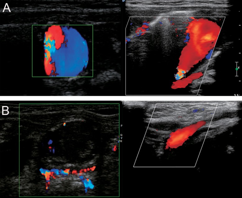
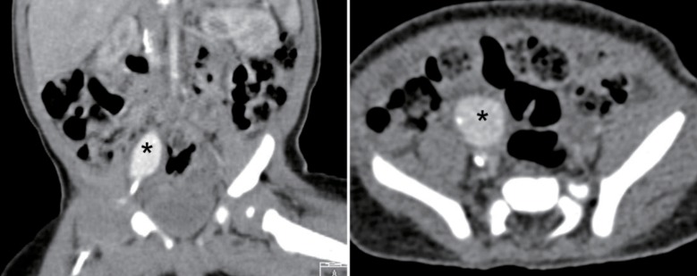
She had elective renal ultrasonography 7 days later to investigate possible renal malformations, as she was suspected to have DiGeorge syndrome with characteristic face and cardiac malformations. Unexpectedly, the ultrasound scan of the kidneys revealed a focal anechoic mass (2.3 cm) with a turbulent flow pattern in the frontal part of the right iliac artery (Fig. 1A). Computed tomography of abdomen clearly demonstrated a pseudoaneurysm with a calcified wall at the anterior aspect of the right iliac vessel (1.6 cm×1.3 cm×2.2 cm), without evidence of arteriovenous fistulas (Fig. 2).
We attempted percutaneous thrombin injection with ultrasound guidance. The infant was sedated with chloral hydrate. Using an attachable biopsy guide, we advanced a 22-gauze Chiba needle tip into the aneurysmal lumen, carefully avoiding pseudoaneurysm neck. After ensuring that the needle tip was optimally positioned at the apex of the pseudoaneurysm, away from the neck, thrombin diluted to 1,000 IU/mL of normal saline was injected slowly under monitoring of the flow in the lumen with color Doppler sonography. The pseudoaneurysm was promptly thromb osed with the injection of 1,000 units of thrombin (Fig. 1B). There were no procedure-related complications. Follow-up ultrasonographic evaluation the next day confirmed complete thrombosis of the pseudoaneurysm without recurrence of perfusion within the former lesion. The patient is doing well with no evidence of complication 20 months after the successful procedure.
Discussion
Several therapeutic methods for the treatment of pseudoaneurysm have been described previously. Traditionally, the standard treatment has been surgical resection. More recently, other less invasive treatment options, such as ultrasound-guided compression of the pseudoaneurysm space, or direct injection of thrombin into the pseudoaneurysm have been employed. In adults, ultrasound-guided thrombin injection has been shown to be more effective than ultrasound-guided compression1).
Thrombin is an enzyme that catalyzes the conversion of fibrinogen to fibrin, and affects the final common pathway in coagulation. Therefore, percutaneous thrombin injection into a confined blood space will lead to a thrombotic cascade. Cope and Zeit5) first described a technique in which thrombin was directly injected into the aneurysmal sac to induce thrombosis and subsequent occlusion of the pseudoaneurysm. The most serious complication of this therapy is embolization of the parent artery. To avoid unintended spillage of thrombin into the parent artery, it is critical to monitor the needle inserted into the pseudoaneurysm to ensure that it is kept away from the neck during the injection.
Although the reported rate of pseudoaneurysms in arterial access sites is up to 8% in adults1), it is rare in children, and information on these lesions in infants or young children is limited.
We applied the ultrasound guided thrombin therapy in a 6-month old infant. To the best of our knowledge, there have been only 2 reports describing successful use of the percutaneous thrombin injection in infants who were 3 weeks and 8 months old, respectively6,7). Although few reports have been published regarding the use of ultrasound-guided thrombin treatment of an iatrogenic pseudoaneurysm in infants, our experience suggests that the technique is safe and effective for the treatment of pseudoaneurysms in such a young age group. We believe that this technique can be considered as the first line treatment for iatrogenic pseudoaneurysm in infants and children.
 PDF Links
 PubReader
 PubMed
 Download Citation


