


Electroencephalography (EEG) is a crucial neuroimaging technique that records brain activity by measuring electrical signals to diagnose neurological disorders, including epilepsy, tumors, and encephalitis [1]. EEG signals are created by the electrical activity induced by the excitation of neurons in the cerebral cortex [2]. Recording electrodes are placed on the scalp to receive electrical input from neurons. Magnetic encephalography (MEG) is another direct imaging technique used to detect electromagnetic signal changes using magnetic sensors placed on the scalp [2]. EEG and MEG can instantly measure electrical or magnetic field changes and offer a high temporal resolution of large-scale brain networks. However, spatial analysis remains restricted [2]. Other imaging modalities have been developed to detect the hemodynamic and metabolic changes in the brain, such as functional magnetic resonance imaging (MRI), positron emission tomography, and single-photon emission computed tomography, which provide good spatial resolution but limited temporal resolution [3].
The localization of active sources in the brain using EEG is referred to as EEG source localization [1]. Source localization of EEG refers to calculating the exact brain location of an electrical current recorded from the scalp [4]. A particular advantage of EEG source localization over other brain imaging methods is its high temporal resolution, which allows us to see the propagation pattern of the origin of activity in brain networks [5]. However, identifying the sources of EEG signal elements in the brain may be difficult because of limited spatial resolution. The development of advanced EEG source localization has resulted in good temporal and spatial resolutions [2]. The introduction of MRI visualized the brain anatomy in detail, while the incorporation of MRI into the source localization of EEG increased its accuracy [5].
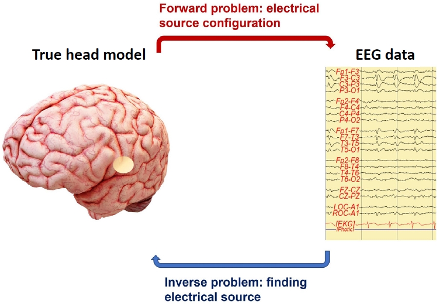
Based on a review by Eom, the source localization process must solve forward and inverse problems (Fig. 1) [1,3,5,6]. The forward problem is the expectation of scalp potentials from current sources and can be solved by an accurate head model [1,3,5]. The shape and conduction distribution of brain tissue strongly influence EEG signals. Therefore, an individualized MRI should be used to construct the exact head model and enable more precise source localization [4]. The inverse problem refers to estimating the precise location of a current source within the brain using scalp potential measurements [1]. One approach to this problem is to set limitations using reasonable assumptions about the volume conductor and generator anatomy. Several proposals have been introduced for the inverse problem [1,2,4,5]. In particular, the author described methods of the source analysis model, such as dipole source localization and distributed source localization. The dipole source localization recorded from scalp EEG can estimate the location sources by calculation of the location, direction, and moment parameters of the current dipoles [4,7]. However, dipole source localization requires an a priori assumption of only a few active areas in the brain, assumes a limited number of equivalent dipoles, and can be biased by missing dipoles [4,5]. Recent developments in brain imaging methods have resulted in more sophisticated options to localize brain sources from scalp EEG signals, and several methods of distributed source localization are currently used [4,5]. The most popular distributed source models are modified algorithms of minimum norm solutions, such as the weighted minimum norm solution, low-resolution electromagnetic tomography, and local autoregressive average [4,5,8].
EEG source localization, a valuable method for investigating brain neural networks and connectivity in clinical and cognitive neuroscience, is often combined with other functional or structural imaging methods [5,9]. The most commonly used methods involve localizing the epileptic focus in focal epilepsy or identifying the eloquent cortex as a presurgical assessment [4]. Furthermore, its clinical application has been extended to other neuropsychiatric diseases such as attention deficit disorder with hyperactivity, anxiety disorder, obsessive-compulsive disorder, and sleep disorders [3,5,10].
In the future, EEG source localization may effectively diagnose several brain disorders. Multimodal methods that combine EEG with high temporal resolution and brain imaging studies with high spatial resolution can provide more helpful information for diagnosing and researching brain abnormalities [2-4]. The applications of EEG source localization are extended by the implementation of advanced technology, such as brain-computer interfaces and machine learning algorithms. EEG source localization using these new technologies will increase our understanding of brain function.
 PDF Links
 PubReader
 ePub Link
 PubMed
 Download Citation


