Warning: fopen(/home/virtual/pediatrics/journal/upload/ip_log/ip_log_2024-04.txt) [function.fopen]: failed to open stream: Permission denied in /home/virtual/pediatrics/journal/ip_info/view_data.php on line 82

Warning: fwrite(): supplied argument is not a valid stream resource in /home/virtual/pediatrics/journal/ip_info/view_data.php on line 83
Diffuse alveolar hemorrhage and recombinant factor VIIa treatment in pediatric patients

Volume 59(3); March

< Previous     Next >

Article Contents

Clin Exp Pediatr > Volume 59(3); 2016
Park: Diffuse alveolar hemorrhage and recombinant factor VIIa treatment in pediatric patients

Abstract

Diffuse alveolar hemorrhage (DAH) is a life-threatening pulmonary complication in patients with hematologic malignancies or autoimmune disorders. The current treatment options, which include corticosteroids, transfusions, extracorporeal membrane oxygenation (ECMO), and immunosuppressants, have been limited and largely unsuccessful. Recombinant activated factor VII (rFVIIa) has been successfully administered, either systemically or bronchoscopically, to adults for the treatment of DAH, but there are few data on its use in pediatric patients. The current literature in the PubMed database was reviewed to evaluate the efficacy and risk of rFVIIa treatment for DAH in pediatric patients. This review discusses the diagnosis and treatment of DAH, as well as a new treatment paradigm that includes rFVIIa. Additionally, the risks and benefits of off-label use of rFVIIa in pediatric patients are discussed.

Introduction

Diffuse alveolar hemorrhage (DAH) is a clinical syndrome that is characterized by alveolar bleeding, which ranges from mild bleeding that requires chronic transfusions to catastrophic bleeding with hemoptysis, pulmonary infiltrates (typically diffuse and bilateral), and hypoxic respiratory failure1,2). The etiology of DAH is diverse and includes pulmonary capillaritis, drugs, toxins, allogeneic hematopoietic stem cell transplantation (HCT), coagulopathies, and blast lung injury. It is a life-threatening condition that is associated with a high mortality rate. However, the treatment of DAH is empirical and ranges from supportive treatment to high-dose corticosteroids, immunosuppressants, and plasmapheresis. Furthermore, the overall mortality remains high.
Recombinant factor VIIa (rFVIIa) was developed for the management of bleeding in hemophiliac patients with inhibitors or in FVII-deficient patients. Despite the narrow indications, the off-label use of rFVIIa has increased among nonhemophiliac patients in order to prevent or treat uncontrolled bleeding in adults, children, and neonates (including preterm neonates)3,4). Recombinant FVIIa has also been reported to have a significant hemostatic effect in patients with DAH5,6,7,8). However, many reports on the use of rFVIIa for DAH were based on adult patients, and few have evaluated rFVIIa use in pediatric patients. This review discusses the diagnosis and etiologies of DAH, the treatment options, and a new treatment paradigm that includes the off-label use of rFVIIa in pediatric patients.

Diagnosis of DAH

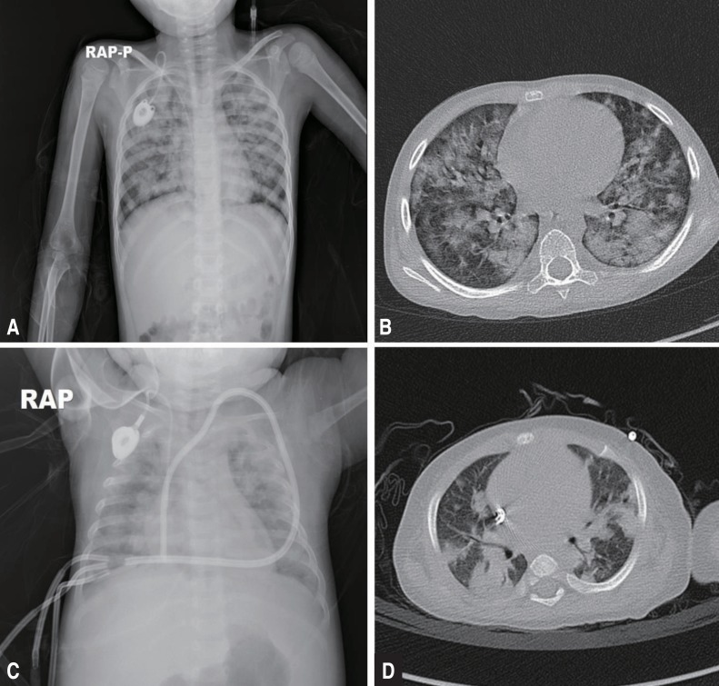
DAHs is characterized by hypoxia, progressive dyspnea, cough, infiltrates on a chest x-ray (CXR), progressively more blood in successive aliquots of bronchoalveolar lavage (BAL) fluid, and the absence of evidence for infectious etiologies in the BAL fluid cultures or other diagnostic studies. Robbins et al.9) first described DAH in 1989. The disease is characterized by damage to the alveolar-capillary basement membrane, which allows red blood cells to enter the alveolar spaces. In adults, DAH is typically a syndrome of pulmonary capillaritis, which is observed with autoimmune diseases or after HCT, and pulmonary hemorrhage after HCT has a high mortality rate10,11). However, DAH may also occur as a consequence of diffuse alveolar damage in patients with acute respiratory distress syndrome/acute lung injury (ARDS/ALI), and can occur in any age group secondary to various underlying diseases and conditions12,13,14,15,16,17). The differential diagnosis is broad, and the etiologies of DAH can be broadly divided according to immune- and non–immune-mediated causes (Table 1)18). A careful history and physical examination can often help to establish the risk factors and most likely etiology of the alveolar hemorrhage. Flexible bronchoscopy is recommended as soon as possible. The diagnosis is established when sequential BAL fluids are progressively more hemorrhagic. Samples should also be sent for routine bacterial, fungal, viral, and Pneumocystis jiroveci studies to rule out infection. The role of open or transbronchial lung biopsies has not been established, but in any child presenting with DAH of unclear cause (particularly after cardiovascular causes have been ruled out), a lung biopsy is strongly recommended to determine the underlying etiology12,19,20). Imaging studies including CXR and high-resolution chest computed tomography scans can provide additional information to support a diagnosis of DAH. However, radiological findings are frequently nonspecific and subject to change throughout the course of the disease. Typical patterns indicative of DAH include focal or diffuse areas of ground glass opacities or consolidations consequent to alveolar filling (Fig. 1). Laboratory studies usually include a complete blood count with differential; coagulation studies; serum blood urea nitrogen and creatinine analysis; antineutrophil cytoplasmic antibody (ANCA) testing (cytoplasmic [c-ANCA] and perinuclear [p-ANCA]); antigen-specific enzyme-linked immunosorbent assay (proteinase 3 or myeloperoxidase-ANCA); analyses of antiglomerular basement membrane antibodies, anti-nuclear antibodies, rheumatoid factor, and antiphospholipid antibodies; urinalysis with urine sediment assessment; and a urine drug screen (Table 2)2).

Treatment of DAH

DAH is a general alveolar response associated with many underlying diseases and conditions. This response can be categorized into 3 subtypes; acute macroscopic bleeding, chronic microscopic alveolar bleeding, and blast lung injury8). DAH, particularly acute macroscopic hemorrhage, has a very high mortality rate; therefore, prompt and aggressive acute management of DAH is critical for survival. It is important to treat the alveolar hemorrhage separately from the underlying diseases, which are treated with plasmapheresis, corticosteroids, immunosuppressants, and antivirals or antibiotics for example (Table 1)8). Acute management of DAH involves supportive care including ventilator support, which ranges from oxygen supplementation to mechanical ventilation with high positive end-expiratory pressure (PEEP). PEEP during mechanical ventilation may produce a tamponade effect to limit capillary bleeding. The coagulation cascade should be evaluated, and any coagulation abnormalities should be corrected. Commonly accepted targets are platelet counts more than 50,000/µL and a prothrombin time-international normalized ratio less than 1.5. Depending on the causes of the coagulation, platelet transfusions, vitamin K supplements, cryoprecipitates, and fresh frozen plasma can be administered for correction.
To control the inflammatory activity, prompt administration of high-dose corticosteroids has been recommended along with treatment for the underlying cause of the hemorrhage. Corticosteroids have been accepted as a mainstay of treatment in most cases, including DAH after HCT, and are aimed at reducing possible acute inflammatory responses to hemorrhage induced by lung alveolar epithelial swelling, thrombotic microangiopathy, and inflammatory cells and cytokines12,20,21). Treatment with high-dose corticosteroids is typically initiated with intravenous methylprednisolone at a dose of 500 mg to 2 g/day or 30 mg/kg/day for the first 3 to 5 days, followed by gradual tapering5,10,20,22). However, these treatments are insufficient to stop acute macroscopic pulmonary bleeding and can be fatal if the etiology is infectious or if the patient is in an immunocompromised state. Despite the widespread use of high-dose corticosteroids for DAH, the overall mortality has remained high; thus, additional treatments for hemostasis are urgently needed.
Traditionally, other treatment modalities for hemostasis have included the use of antifibrinolytics, specifically lysine analogues tranexamic acid (TXA) and epsilon aminocaproic acid (EACA). Although the evidence available for the use of these antifibrinolytics in patients with hematological disorders is limited, they have been used to control perioperative bleeding, mainly in cardiac surgery23,24). In addition to intravenous delivery of TXA, both aerosolized and intrapulmonary administrations have been used to treat DAH and have yielded positive results25). However, a study of TXA in patients with acute promyelocytic leukemia did not reveal any difference in mortality due to bleeding between the patients who received TXA and those who did not26). In addition, some studies have reported an increased risk of postoperative seizures in patients who received TXA, which has since been withdrawn from the market27). An alternative treatment, EACA, which was used in addition to corticosteroids in patients with posttransplant DAH, was reported to result in a lower 100-day mortality rate than treatment with corticosteroids alone28). However, this drug is not available in Korea, and a recent study that included more patients reported that adjuvant treatment with EACA did not yield a difference in the outcomes of patients with DAH after HCT. The overall mortality on day 100 was still high, at 85%29).
In contrast, rFVIIa has generated significant interest and controversy as a potential 'broad-spectrum hemostatic agent' to enhance hemostasis in patients with life-threatening bleeding. Numerous case reports and clinical studies have reported the effectiveness of rFVIIa in various conditions including thrombocytopenia, functional platelet defects, hemorrhagic complications after HCT (e.g., DAH and hemorrhagic cystitis), gunshot wounds, and coagulopathy of liver failure30,31,32,33,34,35,36,37). Whereas there is overwhelming evidence that rFVIIa has hemostatic effects in nonhemophiliac patients, information on the efficacy and side effects of rFVIIa in nonhemophiliac settings is largely anecdotal. Furthermore, many questions remain regarding the appropriate settings and guidelines for use; risks of thrombotic complications; and appropriate monitoring, dosing, and integration with transfusion therapy. A recent Cochrane review regarding the off-label use of rFVIIa concluded that therapeutic use of rFVIIa did not significantly decrease mortality compared with placebo (odds ratio [OR], 0.91; 95% confidence interval [CI], 0.78–1.06), nor did it significantly increase the incidence of thromboembolic events (OR, 1.18; 95% CI, 0.94–1.48). However, there was a trend towards control of the bleeding, and a lower number of transfusions favoring rFVIIa (OR, 0.95; 95% CI, 0.88–1.03; and OR, 0.94; 95% CI, 0.89–1.00), whereas there was a significant increase in arterial thromboembolic events (OR. 1.45; 95% CI. 1.02–2.05)38). Because this meta-analysis included a wide range of disorders (from HCT to trauma to hemorrhagic stroke), a significant effect on the outcome in one clinical setting might have been diminished by a lack of benefit in another. There are many anecdotal reports on the successful use of rFVIIa in patients with DAH following various diseases and injuries including pneumonia, HCT, metastatic cancer, idiopathic pulmonary hemosiderosis, and immune-associated vasculitis; therefore, the use of rFVIIa for the treatment of DAH deserves consideration.

rFVIIa treatment for DAH

There are an increasing number of studies in the literature supporting the use of rFVIIa to stop acute pulmonary hemorrhage. Due to the lack of a randomized controlled study, rFVIIa has been used in patients with severe pulmonary hemorrhage as a final effort to stop the bleeding. Recently, several studies have reported the successful use of rFVIIa for life-threatening DAH in adults, as well as in children and infants6,13,39,40,41,42). In refractory cases of immune- and non–immune-mediated DAH, including cases due to pulmonary vasculitis, connective tissue diseases, post-HCT, or infections, rFVIIa has been successfully administered both systemically (intravenously) and bronchoscopically. Although the optimal dose and dosing intervals remain to be determined, systemic administration usually involves the intravenous administration of 35–200 µg/kg as either a single dose or repeated doses every 2–4 hours5,7,40,43). Intrapulmonary therapy typically involves bronchoscopy, with a total dose of 50–90 µg/kg of rFVIIa diluted in normal saline and administered as either a single dose or, if bleeding continues, as repeated doses over 24 hours 8,13,44,45).
Recombinant FVIIa promotes hemostasis via both a tissue factor (TF)-dependent pathway at the sites of endothelial injury and a TF-independent pathway, directly activating factors IX and X on the surface of activated platelets in the absence of TF and at higher doses. Factor X subsequently converts prothrombin to thrombin, which in turn converts fibrinogen to fibrin. The alveolar TF concentration remains high in inflammatory pulmonary conditions including DAH, ARDS, and pneumonia, as well as after local lipopolysaccharide challenge in the alveoli6,46,47,48). The alveolar TF-rFVIIa complex activates coagulation factors IX and X, which in turn initiate a cascade of reactions, leading to thrombin burst and fibrin formation at the site of injury6). Given the separation between the alveolar and systemic compartments of the lung, rFVIIa requires a high systemic concentration to affect specific receptors in the alveolar compartment, similar to most biologics. This effect necessitates higher and repeated doses of rFVIIa, which may be associated with a higher risk of thromboembolic complications8,49,50). Recently, Pathak et al.42) reported that bleeding resolved in 22 of 23 DAH patients after systemically administered rFVIIa treatment, without adverse thrombotic events. However, the mean dose of rFVIIa required to control the bleeding was 5±3 mg, and the authors emphasized that an adequate antithrombin III level should be maintained during rFVIIa treatment to avoid the generation of thrombosis, an adverse effect7).
Meanwhile, successful outcomes have been reported for patients with DAH after treatment with intrapulmonary rFVIIa at a relatively low dose (50 µg/kg)6,13,41). Local administration of rFVIIa into the airways can ensure that it reaches the alveolar receptor (TF), and it may not have systemic adverse effects because the alveolocapillary membrane does not allow the transmembranous passage of rFVIIa. Pulmonary-administered rFVIIa combines with TF and can lead to balanced hemostasis, with a small dose of rFVIIa resulting in both durable hemostasis and a significant improvement in oxygen transport capacity. Moreover, the TF pathway inhibitor, an inhibitor of the local activation of factor X that is produced by alveolar macrophages, is highly expressed in the airspace in inflammatory conditions secondary to ALI. This characteristic may contribute to balanced hemostasis without the theoretical risk of intra-alveolar thrombotic complications when treating DAH with intrapulmonary rFVIIa8,51,52). Accordingly, the treatment with rFVIIa has a potentially high benefit-to-risk ratio in DAH when administered via the local intrapulmonary route.

rFVIIa treatment in pediatric patients

The off-label use of rFVIIa in pediatric patients has also increased despite the absence of adequate studies addressing safety and efficacy. According to a multicenter cohort study by Witmer et al.53), 83% of rFVIIa use was off-label, and the off-label use of rFVIIa increased 10 folds between 2000 and 2007. The Haemostasis Registry reported the results of off-label use of rFVIIa in 388 pediatric patients54). The median age was 12 months, and the median dosages (single and total doses) were 114 µg/kg and 1.2 mg, respectively. A reduction in the number of transfusions after the use of rFVIIa (P<0.01) was observed, and the subjective response rate was 82%. In both studies, thromboembolic complications were reported in 5.4% to 10.9% of patients, which were similar to the published rates for adults49,55,56). The results demonstrated that rFVIIa was an effective and safe hemostatic agent in severely ill children experiencing life-threatening hemorrhages and that rFVIIa, when administered along with standard treatment modalities, reduced blood product requirements in children3,53,54,57). However, the benefits of rFVIIa therapy in neonates and infants less than one year of age remain unclear. Young et al.57) reported that neonates had a lower response rate (47%) and a higher rate of thrombosis (17.6%) than older age groups (greater than one month of age). The SeveNBleeP registry (a Web-based registry of rFVIIa use in nonhemophiliac children) of 42 infants less than 1 year of age also revealed no significant reduction in the requirements for blood products after rFVIIa treatment and indicated that there was no significant increase in thromboembolism (2.4%)3). There is evidence that rFVIIa has a shorter half-life and more rapid clearance in children compared to adults; therefore, higher and more frequent doses of rFVIIa are often required to control significant bleeding in children, especially in neonates and infants less than 1 year of age3,54,57). Considering the relatively poor response to rFVIIa and that the levels of natural inhibitors of coagulation (proteins C and S and antithrombin-III) are reduced, the risk of thromboembolism can increase in neonates and infants, and the risk-to-benefit ratio can be higher. Thus, caution has been recommended when considering treatment of patients in this age group with rFVIIa.
Perinatal asphyxia, very low birth weight, mechanical ventilation, respiratory distress syndrome, exogenous surfactant therapy, sepsis, hypothermia, patent ductus arteriosus, and coagulopathies can also be causes of pulmonary hemorrhage in children including neonates4,58). There are few reports regarding the treatment of pediatric patients with rFVIIa for pulmonary hemorrhage (Table 3). The data for 37 patients have been published, and the outcome demonstrated a complete response (bleeding completely ceased) in 62% and a partial response (bleeding improved but did not cease) in 24% of the patients. No response to rFVIIa treatment was observed for 13.5% of the patients. Two thromboembolic complications were reported (5.4%)57,59). Twenty-nine patients received intravenous rFVIIa, with a median total dose of 175 µg/kg (range, 60–3,150 µg/kg), and satisfactory hemostasis was achieved through multiple doses of rFVIIa in most cases. Complete hemostasis was reported in 14 cases (48%), a partial response in 10 cases (35%), and no response in 5 cases (17%). Although these data were not the representative of the effect of rFVIIa for DAH in pediatric patients, intravenous rFVIIa appeared to be less effective for the treatment of pulmonary hemorrhage compared to other life-threatening hemorrhages in children, and this finding was consistent with those of previous studies40,57). It is possible that because of pharmacokinetic characteristics, higher and multiple doses of rFVIIa may be necessary to reach an adequate systemic concentration to affect TFs in the alveolar compartment of the lung and to achieve stable hemostasis in children; however, the potential risk for thromboembolic complications also increases concomitantly.
Following the report of Heslet et al.6), several case series have been reported the use of intrapulmonary rFVIIa for the treatment of DAH in pediatric patients. Eight patients were treated with intrapulmonary rFVIIa, and complete hemostasis was observed in all cases with a median dose of 50 µg/kg. Most patients showed a significant improvement in oxygenation capacity (PaO2/FiO2 ratio) on the days following rFVIIa treatment13,60), although one child experienced a thrombotic obstruction in the endotracheal tube (ETT)59). There was no evidence of intra-alveolar thrombotic deposition (hyaline membrane formation) after rFVIIa administration among the eight pediatric patients. The data also suggested that pulmonary hemostasis could be more easily induced from the alveolar side than from the endothelial side, and that the intrapulmonary administration of rFVIIa could be more effective for the treatment of DAH in both children and adults.
Collectively, the data have suggested that intrapulmonary administration of rFVIIa was effective in controlling DAH in pediatric patients who have a lower systemic risk of complications; however, the risk of critical airway or ETT obstruction in young patients should be carefully considered.

Conclusions

DAH should be suspected in any patient who presents with alveolar infiltrates on CXR, hypoxemia, anemia, and hemoptysis. It is a clinical syndrome that can be a manifestation of multiple different etiologies, and identification of the underlying etiology is important for determining the treatment options. In cases of life-threatening DAH, rFVIIa therapy along with the appropriate treatment for the underlying disease may contribute to patient survival. Although most of existing data are based on case series and reports; thus, the results could have been affected by selection bias, the available data have demonstrated the safety and efficacy of rFVIIa therapy in uncontrolled nonhemophiliac bleedings in pediatric patients. The limiting factors in its use include the high cost ($1,000 per gram) and the substantial risk for thromboembolic complications. In this regard, the intrapulmonary administration of a low dose of rFVIIa may be an effective and reasonable treatment option for DAH in children as well as in adults. However, without the appropriate treatment of the primary causes of DAH, rFVIIa therapy would only functions as a temporary solution. Additional data from prospective registries or randomized trials when feasible are necessary to further our knowledge regarding the use of rFVIIa in pediatric patients with DAH.

Conflicts of interest

Conflicts of interest:
No potential conflict of interest relevant to this article was reported.

References

1. Collard HR, Schwarz MI. Diffuse alveolar hemorrhage. Clin Chest Med 2004;25:583–592.
crossref pmid
2. Lara AR, Schwarz MI. Diffuse alveolar hemorrhage. Chest 2010;137:1164–1171.
crossref pmid
3. Blatny J, Mathew P, Monagle P, Ovesna P, Fiamoli V. Safety and efficacy of recombinant activated factor VII in nonhemophilia children with severe or life-threatening bleeding: a report from the SeveNBleeP registry. Blood Coagul Fibrinolysis 2014;25:326–332.
crossref pmid
4. Cetin H, Yalaz M, Akisu M, Karapinar DY, Kavakli K, Kultursay N. The use of recombinant activated factor VII in the treatment of massive pulmonary hemorrhage in a preterm infant. Blood Coagul Fibrinolysis 2006;17:213–216.
crossref pmid
5. Pastores SM, Papadopoulos E, Voigt L, Halpern NA. Diffuse alveolar hemorrhage after allogeneic hematopoietic stem-cell transplantation: treatment with recombinant factor VIIa. Chest 2003;124:2400–2403.
crossref pmid
6. Heslet L, Nielsen JD, Levi M, Sengelov H, Johansson PI. Successful pulmonary administration of activated recombinant factor VII in diffuse alveolar hemorrhage. Crit Care 2006;10:R177
crossref pmid pmc
7. Henke D, Falk RJ, Gabriel DA. Successful treatment of diffuse alveolar hemorrhage with activated factor VII. Ann Intern Med 2004;140:493–494.
crossref pmid
8. Heslet L, Nielsen JD, Nepper-Christensen S. Local pulmonary administration of factor VIIa (rFVIIa) in diffuse alveolar hemorrhage (DAH) - a review of a new treatment paradigm. Biologics 2012;6:37–46.
pmid pmc
9. Robbins RA, Linder J, Stahl MG, Thompson AB 3rd, Haire W, Kessinger A, et al. Diffuse alveolar hemorrhage in autologous bone marrow transplant recipients. Am J Med 1989;87:511–518.
crossref pmid
10. Majhail NS, Parks K, Defor TE, Weisdorf DJ. Diffuse alveolar hemorrhage and infection-associated alveolar hemorrhage following hematopoietic stem cell transplantation: related and high-risk clinical syndromes. Biol Blood Marrow Transplant 2006;12:1038–1046.
crossref pmid
11. Afessa B, Tefferi A, Litzow MR, Krowka MJ, Wylam ME, Peters SG. Diffuse alveolar hemorrhage in hematopoietic stem cell transplant recipients. Am J Respir Crit Care Med 2002;166:641–645.
crossref pmid
12. Newsome BR, Morales JE. Diffuse alveolar hemorrhage. South Med J 2011;104:269–274.
crossref pmid
13. Park JA, Kim BJ. Intrapulmonary recombinant factor VIIa for diffuse alveolar hemorrhage in children. Pediatrics 2015;135:e216–e220.
crossref pmid
14. Lee DS, Lee YI, Ahn JB, Kim MJ, Kim JH, Kim NH, et al. Massive pulmonary hemorrhage in enterovirus 71-infected hand, foot, and mouth disease. Korean J Pediatr 2015;58:112–115.
crossref pmid pmc
15. Lee JY, Chon GR, Park JH, Kang BJ, Shim TS, Jo KW. Massive hemoptysis due to pulmonary vein stenosis following catheter ablation for atrial fibrillation. Respir Care 2015;60:e52–e55.
crossref pmid
16. Fouret PJ, Touboul JL, Mayaud CM, Akoun GM, Roland J. Pulmonary Kaposi's sarcoma in patients with acquired immune deficiency syndrome: a clinicopathological study. Thorax 1987;42:262–268.
crossref pmid pmc
17. Kim JS, Kim MJ, Bae EY, Jeong DC. Pulmonary hemorrhage in pediatric lupus anticoagulant hypoprothrombinemia syndrome. Korean J Pediatr 2014;57:202–205.
crossref pmid pmc
18. Krause ML, Cartin-Ceba R, Specks U, Peikert T. Update on diffuse alveolar hemorrhage and pulmonary vasculitis. Immunol Allergy Clin North Am 2012;32:587–600.
crossref pmid pmc
19. Travis WD, Colby TV, Lombard C, Carpenter HA. A clinicopathologic study of 34 cases of diffuse pulmonary hemorrhage with lung biopsy confirmation. Am J Surg Pathol 1990;14:1112–1125.
crossref pmid
20. Susarla SC, Fan LL. Diffuse alveolar hemorrhage syndromes in children. Curr Opin Pediatr 2007;19:314–320.
crossref pmid
21. Heggen J, West C, Olson E, Olson T, Teague G, Fortenberry J, et al. Diffuse alveolar hemorrhage in pediatric hematopoietic cell transplant patients. Pediatrics 2002;109:965–971.
crossref pmid
22. Raptis A, Mavroudis D, Suffredini A, Molldrem J, Rhee FV, Childs R, et al. High-dose corticosteroid therapy for diffuse alveolar hemorrhage in allogeneic bone marrow stem cell transplant recipients. Bone Marrow Transplant 1999;24:879–883.
crossref pmid
23. Sindet-Pedersen S, Ramström G, Bernvil S, Blombäck M. Hemostatic effect of tranexamic acid mouthwash in anticoagulant-treated patients undergoing oral surgery. N Engl J Med 1989;320:840–843.
crossref pmid
24. Taghaddomi RJ, Mirzaee A, Attar AS, Shirdel A. Tranexamic acid reduces blood loss in off-pump coronary artery bypass surgery. J Cardiothorac Vasc Anesth 2009;23:312–315.
crossref pmid
25. Solomonov A, Fruchter O, Zuckerman T, Brenner B, Yigla M. Pulmonary hemorrhage: a novel mode of therapy. Respir Med 2009;103:1196–1200.
crossref pmid
26. Sanz MA, Montesinos P. Open issues on bleeding and thrombosis in acute promyelocytic leukemia. Thromb Res 2010;125(Suppl 2): S51–S54.
crossref pmid
27. Sander M, Spies CD, Martiny V, Rosenthal C, Wernecke KD, von Heymann C. Mortality associated with administration of high-dose tranexamic acid and aprotinin in primary open-heart procedures: a retrospective analysis. Crit Care 2010;14:R148
crossref pmid pmc
28. Wanko SO, Broadwater G, Folz RJ, Chao NJ. Diffuse alveolar hemorrhage: retrospective review of clinical outcome in allogeneic transplant recipients treated with aminocaproic acid. Biol Blood Marrow Transplant 2006;12:949–953.
crossref pmid
29. Rathi NK, Tanner AR, Dinh A, Dong W, Feng L, Ensor J, et al. Low-, medium- and high-dose steroids with or without aminocaproic acid in adult hematopoietic SCT patients with diffuse alveolar hemorrhage. Bone Marrow Transplant 2015;50:420–426.
crossref pmid
30. Kristensen J, Killander A, Hippe E, Helleberg C, Ellegard J, Holm M, et al. Clinical experience with recombinant factor VIIa in patients with thrombocytopenia. Haemostasis 1996;26(Suppl 1): 159–164.
pmid
31. Al Hammadi AM, Sallah S. Efficacy and safety of recombinant factor VIIa in the treatment of bleeding episodes in patients with aplastic anemia. J Thromb Haemost 2007;5:435–436.
crossref pmid
32. Farah RA, Hamod D, Melick N, Giansily-Blaizot M, Sallah S. Successful prophylaxis against intracranial hemorrhage using weekly administration of activated recombinant factor VII in a newborn with severe factor VII deficiency. J Thromb Haemost 2007;5:433–434.
crossref pmid
33. Tengborn L, Petruson B. A patient with Glanzmann thrombasthenia and epistaxis successfully treated with recombinant factor VIIa. Thromb Haemost 1996;75:981–982.
crossref
34. Kenet G, Walden R, Eldad A, Martinowitz U. Treatment of traumatic bleeding with recombinant factor VIIa. Lancet 1999;354:1879
crossref pmid
35. Pihusch M, Bacigalupo A, Szer J, von Depka Prondzinski M, Gaspar-Blaudschun B, Hyveled L, et al. Recombinant activated factor VII in treatment of bleeding complications following hematopoietic stem cell transplantation. J Thromb Haemost 2005;3:1935–1944.
crossref pmid
36. Yadav SP, Sachdeva A, Bhat S, Katewa S. Successful control of massive gastrointestinal bleeding following umbilical cord blood transplantation (UCBT) by use of recombinant activated factor VII (rFVIIa) and octreotide infusion. Pediatr Hematol Oncol 2010;27:24–30.
crossref pmid
37. Harkensee C, Vasdev N, Gennery AR, Willetts IE, Taylor C. Prevention and management of BK-virus associated haemorrhagic cystitis in children following haematopoietic stem cell transplantation: a systematic review and evidence-based guidance for clinical management. Br J Haematol 2008;142:717–731.
crossref pmid
38. Simpson E, Lin Y, Stanworth S, Birchall J, Doree C, Hyde C. Recombinant factor VIIa for the prevention and treatment of bleeding in patients without haemophilia. Cochrane Database Syst Rev 2012;3:CD005011

39. Brady KM, Easley RB, Tobias JD. Recombinant activated factor VII (rFVIIa) treatment in infants with hemorrhage. Paediatr Anaesth 2006;16:1042–1046.
crossref pmid
40. Bhat S, Yadav SP, Anjan M, Dinand V, Sachdeva A. Recombinant activated factor VII usage in life threatening hemorrhage: a pediatric experience. Indian J Pediatr 2011;78:961–968.
crossref pmid
41. Esper RC, Estrada IE, de la Torre Leon T, Gutiérrez AO, Lopez JA. Treatment of diffuse alveolar hemorrhage secondary to lupus erythematosus with recombinant activated factor VII administered with a jet nebulizer. J Intensive Care 2014;2:47
crossref pmid pmc
42. Pathak V, Kuhn J, Gabriel D, Barrow J, Jennette JC, Henke DC. Use of activated factor VII in patients with diffuse alveolar hemorrhage: a 10 years institutional experience. Lung 2015;193:375–379.
crossref pmid
43. Elinoff JM, Bagci U, Moriyama B, Dreiling JL, Foster B, Gormley NJ, et al. Recombinant human factor VIIa for alveolar hemorrhage following allogeneic stem cell transplantation. Biol Blood Marrow Transplant 2014;20:969–978.
crossref pmid pmc
44. Dabar G, Harmouche C, Jammal M. Efficacy of recombinant activated factor VII in diffuse alveolar haemorrhage. Rev Mal Respir 2011;28:106–111.
crossref pmid
45. Estella A, Jareno A, Perez-Bello Fontaina L. Intrapulmonary administration of recombinant activated factor VII in diffuse alveolar haemorrhage: a report of two case stories. Cases J 2008;1:150
crossref pmid pmc
46. Schultz MJ, Haitsma JJ, Zhang H, Slutsky AS. Pulmonary coagulopathy as a new target in therapeutic studies of acute lung injury or pneumonia: a review. Crit Care Med 2006;34:871–877.
crossref pmid
47. Schultz MJ, Millo J, Levi M, Hack CE, Weverling GJ, Garrard CS, et al. Local activation of coagulation and inhibition of fibrinolysis in the lung during ventilator associated pneumonia. Thorax 2004;59:130–135.
crossref pmid pmc
48. Levi M, Schultz MJ, Rijneveld AW, van der Poll T. Bronchoalveolar coagulation and fibrinolysis in endotoxemia and pneumonia. Crit Care Med 2003;31(4 Suppl): S238–S242.
crossref pmid
49. Levi M, Levy JH, Andersen HF, Truloff D. Safety of recombinant activated factor VII in randomized clinical trials. N Engl J Med 2010;363:1791–1800.
crossref pmid
50. Stanworth SJ, Birchall J, Doree CJ, Hyde C. Recombinant factor VIIa for the prevention and treatment of bleeding in patients without haemophilia. Cochrane Database Syst Rev 2007;(2): CD005011
pmid
51. Ellery PE, Adams MJ. Tissue factor pathway inhibitor: then and now. Semin Thromb Hemost 2014;40:881–886.
crossref pmid
52. Wood JP, Ellery PE, Maroney SA, Mast AE. Biology of tissue factor pathway inhibitor. Blood 2014;123:2934–2943.
crossref pmid pmc
53. Witmer CM, Huang YS, Lynch K, Raffini LJ, Shah SS. Off-label recombinant factor VIIa use and thrombosis in children: a multi-center cohort study. J Pediatr 2011;158:820–825.e1.
crossref pmid
54. McQuilten ZK, Barnes C, Zatta A, Phillips LE. Haemostasis Registry Steering Committee. Off-label use of recombinant factor VIIa in pediatric patients. Pediatrics 2012;129:e1533–e1540.
crossref pmid
55. MacLaren R, Weber LA, Brake H, Gardner MA, Tanzi M. A multicenter assessment of recombinant factor VIIa off-label usage: clinical experiences and associated outcomes. Transfusion 2005;45:1434–1442.
crossref pmid
56. O'Connell KA, Wood JJ, Wise RP, Lozier JN, Braun MM. Thromboembolic adverse events after use of recombinant human coagulation factor VIIa. JAMA 2006;295:293–298.
crossref pmid
57. Young G, Wicklund B, Neff P, Johnson C, Nugent DJ. Off-label use of rFVIIa in children with excessive bleeding: a consecutive study of 153 off-label uses in 139 children. Pediatr Blood Cancer 2009;53:179–183.
crossref pmid
58. Fekete M, Nemeth A. Neonatal pulmonary haemorrhage, birthweight, gestational age and intrauterine growth. Acta Paediatr Hung 1985;26:65–73.
pmid
59. Larcombe PJ, Kapur N, Fraser CJ, Coulthard MG, Schlapbach LJ. Intrabronchial administration of activated recombinant factor VII in a young child with diffuse alveolar hemorrhage. Pediatr Blood Cancer 2014;61:570–571.
crossref pmid
60. Colin AA, Shafieian M, Andreansky M. Bronchoscopic instillation of activated recombinant factor VII to treat diffuse alveolar hemorrhage in a child. Pediatr Pulmonol 2010;45:411
crossref pmid
61. Blatt J, Gold SH, Wiley JM, Monahan PE, Cooper HC, Harvey D. Off-label use of recombinant factor VIIa in patients following bone marrow transplantation. Bone Marrow Transplant 2001;28:405–407.
crossref pmid
62. Veldman A, Fischer D, Voigt B, Beyer PA, Schlosser R, Allendorf A, et al. Life-threatening hemorrhage in neonates: management with recombinant activated factor VII. Intensive Care Med 2002;28:1635–1637.
crossref pmid
63. Olomu N, Kulkarni R, Manco-Johnson M. Treatment of severe pulmonary hemorrhage with activated recombinant factor VII (rFVIIa) in very low birth weight infants. J Perinatol 2002;22:672–674.
crossref pmid
64. Leibovitch L, Kenet G, Mazor K, Matok I, Vardi A, Barzilay Z, et al. Recombinant activated factor VII for life-threatening pulmonary hemorrhage after pediatric cardiac surgery. Pediatr Crit Care Med 2003;4:444–446.
crossref pmid
65. Grizelj R, Vukovic J, Filipovic-Grcic B, Saric D, Luetic T. Successful use of recombinant activated FVII and aminocaproic acid in four neonates with life-threatening hemorrhage. Blood Coagul Fibrinolysis 2006;17:413–415.
crossref pmid
Fig. 1

Chest radiographs (A, C) and high-resolution computed tomography scans (B, D) showing bilateral patch infiltrates and diffuse ground glass opacities in both lung fields.

kjped-59-105-g001.jpg
Table 1

The etiologies of diffuse alveolar hemorrhage and the treatment options

kjped-59-105-i001.jpg
Clinical presentation Treatment
Immune mediated
 ANCA-associated vasculitis
  Granulomatosis with polyangitis Immunosuppressants
  Microscopic polyangitis Corticosteroids
  Eosinophilic granulomatosis with polyangitis Methotrexate
 Systemic lupus erythematosus Cyclophosphamide
 Rheumatoid arthritis Rituximab
 Inflammatory myopathies Azathioprine
 Antiphospholipid antibody syndrome
 Henoch-Schönlein purpura IgG
 IgA nephropathy
 Cryoglobulinemia Removal of autoantibodies
 Behçet disease Plasmapheresis
 Hypocomplementemic urticarial vasculitis (anti-C1q vasculitis)
 Lung transplant rejection
 Drug-induced vasculitis
 Medications: warfarin, aspirin, amiodarone, phenytoin Withdrawal of offending medications
 Anti-GBM antibody syndrome (Goodpasture's syndrome)
 Idiopathic pulmonary capillaritis
Nonimmune mediated
 Cardiovascular disease
  Mitral stenosis Procoagulation
  Arteriovenous malformation Fresh frozen plasma
  Pulmonary SOS Tranexamic acid
  Pulmonary lymphangiomyomatosis Platelet transfusion
  Pulmonary hypertension Cryoprecipitates
  Pulmonary capillary hemangiomatosis Vitamin K supplementation
  Left ventricular dysfunction
 Noncardiovascular disease
  Infection
  Pseudomonas aeruginosa Broad-spectrum antibiotics
  Aspergillus spp. Antifungal agents
  CMV and herpes pneumonitis Antiviral therapy
 Diffuse alveolar damage
  Radiation
  Cytotoxic drugs
  Acute respiratory distress syndrome
 Hematopoietic stem cell transplantation
 Idiopathic pulmonary hemosiderosis
 Acute idiopathic pulmonary hemorrhage of infancy
 Heiner syndrome (cow's milk hypersensitivity)
 Coagulopathy

ANCA, antineutrophil cytoplasmic antibodies; C1q, complement 1q; CMV, cytomegalovirus; GBM, glomerular basement membrane.

Table 2

Evaluation of diffuse alveolar hemorrhage

kjped-59-105-i002.jpg
Nonspecific findings Etiologies
Laboratory
 Leukocytosis Any cause
 Thrombocytopenia Cytotoxic drugs
Antiphospholipid syndrome
SLE
 Increased ESR Any cause
 Urine RBC casts Any systemic vasculitis
SLE, mixed connective tissue disease
Anti-GBM disease
 Prolonged PT/aPTT/PT INR SLE
Coagulation disorders, DIC, sepsis
Drugs
Radiological
 Patch diffuse alveolar infiltrates Any cause
 Air bronchogram Any cause
PFT
 Obstructive pattern Acute period
 Restrictive pattern Chronic period
Specific findings
 Laboratory
  ANA Connective tissue disease
  RF Connective tissue disease
ANCA-associated granulomatous vasculitis
  c-ANCA Granulomatosis with polyangitis (Wegener's granulomatosis)
Microscopic polyangitis
  p-ANCA Microscopic polyangitis
  Anti-MPO ANCA-associated granulomatous vasculitis
Eosinophilic granulomatosis with polyangitis (Churg-Straus syndrome)
  Anti-GBM antibody Goodpasture syndrome (anti-GBM disease)
 Radiological
  Kerley B lines Pulmonary SOS, MS
  Nodules, cavities Wegener granulomatosis
Rheumatoid arthritis

SLE, systemic lupus erythematosus; ESR, erythrocyte sedimentation rate; RBC, red blood cell; anti-GBM, anti-glomerular basement membrane; PT, prothrombin time; aPTT, activated partial thromboplastin time; INR, international normalized ratio; DIC, disseminated intravascular coagulation; PFT, pulmonary function test; ANA, anti-nuclear antibody; RF, rheumatoid factor; c-ANCA, cytoplasmic anti-neutrophil cytoplasmic antibodies; p-ANCA, perinuclear anti-neutrophil cytoplasmic antibodies; anti-MPO, anti-myeloperoxidase; SOS, sinusoidal obstruction syndrome; MS, mitral stenosis.

Table 3

Published data on recombinant factor VIIa treatment for diffuse alveolar hemorrhage in children

kjped-59-105-i003.jpg
Publication year Patients (sex/age) Primary diagnosis Previous history before DAH Route Total dose of rFVIIa (g/kg) rFVIIa effect Complication Rebleeding Survival Reference
2001 F/8 yr AML Post-HCT, HC IV 3,150 PR No Yes No 61
2002 M/preterm VLBW, RDS, PDA, IVH PDA ligation IV 400 CR No No Yes 62
2002 M/preterm VLBW, RDS, PDA Sepsis, DIC, PIE IV 180 CR No No Yes 63
F/preterm VLBW, RDS, PDA Sepsis, DIC IV 600 CR No No Yes
2003 F/2 mo Down syndrome, CHD Cardiac surgery IV 400 CR No No Yes 64
2006 M/preterm LBW, RDS Sepsis, DIC IV 540 PR No No Yes 4
2006 F/2 days MMA DIC, HD for hyperammonemia IV 180 CR No No Yes 39
M/2 days No Pseudomonal sepsis IV 90 CR No No No
2006 NA/0 day MAS Ventilator care IV 170 CR No No Yes 65
NA/13 days HLHS Postsurgical resuscitation IV 523 CR No No Yes
NA/2 days HLHS Postsurgical resuscitation IV 444 CR No No Yes
2009 12 Patients NA NA IV 90 (range, 20.3–353) 4 CR, 5 PR, 3 NR 1 LV thrombus - NA 57
2011 M/14 yr DSS Sepsis IV 70 CR No No Yes 40
M/13 yr AML TLS, ARF, acute pancreatitis IV 90 PR No - No
F/9 yr Thalassemia, major Post-HCT, ARDS IV 90 CR No - Yes
F/13 yr ALL Febrile neutropenia, sepsis IV 70 NR No - No
M/10 yr AML None IV 75 NR No - No
F/6 yr IPH Pneumothorax and septic shock IV 60 PR No - No
2010 M/17 yr AML Pancytopenia, sepsis IP 50 CR No No Yes 60
2014 M/2 yr AML Post-HCT, hepatic SOS, GVHD IP 50 CR ETT thrombus Yes No 59
2015 F/11 yr MDS Post-HCT, HC, TMA IP 60 CR No No No 13
M/15 yr AML DIC, cytarabine syndrome IP 45 CR No No Yes
M/6 yr T-LL Chickenpox infection, hepatic SOS IP 43 CR No No Yes
M/14 yr AML DIC IP 52 CR No No Yes
F/11 yr IPH None IP 57 CR No No Yes
F/10 mo HLH Post-HCT, CMV infection, hepatic SOS IP 63 CR No No No

rFVIIa, recombinant factor VIIa; DAH, diffuse alveolar hemorrhage; AML, acute myeloid leukemia; HCT, hematopoietic stem cell transplantation; HC, hemorrhagic cystitis; IV, intravenously; PR, partial response; VLBW, very low birth weight; RDS, respiratory distress syndrome; PDA, patent ductus arteriosus; IVH, intraventricular hemorrhage; CR, complete response; DIC, disseminated intravascular coagulation; PIE, pulmonary interstitial emphysema; CHD, complex heart disease; LBW, low birth weight; MMA, methylmalonic aciduria; HD, hemodialysis; MAS, meconium aspiration syndrome; HLHS, hypoplastic left heart syndrome; NA, not available; NR, no response; LV, left ventricle; DSS, dengue shock syndrome; TLS, tumor lysis syndrome; ARF, acute renal failure; ARDS, acute respiratory distress syndrome; ALL, acute lymphoblastic leukemia; IPH, idiopathic pulmonary hemosiderosis; IP, intrapulmonary; SOS, sinusoidal obstruction syndrome; GVHD, graft-versus-host disease; ETT, endotracheal tube; MDS, myelodysplastic syndrome; TMA, thrombotic microangiopathy; T-LL, T-cell lymphoblastic lymphoma; HLH, hemophagocytic lymphohistiocytosis; CMV, cytomegalovirus; No, no underlying disease.

METRICS Graph View
  • 5 Crossref
  • 7 Scopus
  • 10,667 View
  • 232 Download