

Introduction
Congenital syphilis, caused by Treponema pallidum is still a public health issue worldwide, especially in developing countries1). Accordingly, it is important to be familiar with its symptoms, which can be subtle and nonspecific2). Recently, an increase in the incidence and prevalence of early congenital syphilis was observed in Korea as well as other Western countries despite prenatal serologic screening and treatment programs which are included in the routine antenatal examination1,3). Therefore, physicians should be aware of the diverse clinical features of syphilis to enable early diagnosis of the disease. We report here a case of congenital syphilis in a 3-month-old infant who had whole body skin eruption and no other specific symptoms, which led to a delay in diagnosis. His mother was tested for the disease during the prenatal period and the test was negative; however, she tested positive for syphilis later.
Case report
A 3-month-old boy was admitted due to a 3-week history of an asymptomatic, widespread skin eruption. He was given the Diphtheria-Tetanus-acellular Pertussis (DTaP) vaccination 2 days prior to skin rash and his parents had taken him to several pediatric and dermatologic clinics, but could not identify his illness. He had been born to a 24-year-old, G1P1A0 mother who had adequate prenatal care at other clinics. She was tested for syphilis at 12 and 38 weeks of gestational age and was told that the tests were negative. The patient was delivered at 39+6 weeks of gestational age by normal spontaneous vaginal delivery and the birth weight was 3,270 g.
Upon physical examination, his height was 61.5 cm (25 to 50th percentile), weight was 6.5 kg (25 to 50th percentile), and head circumference was 39.5 cm (10 to 25th percentile). He ate only a small amount of milk and his activity was decreased. His abdomen was soft and liver and spleen were not palpable. He had generalized erythematous, targetoid, scaly macules, papules, pustules (Fig. 1A) and desquamation at the hand and foot (Fig. 1B).
Complete blood cell counts demonstrated normocytic normochromic anemia (hemoglobin 8.3 g/dL and hematocrit 27.8%) with leukocytosis (white blood cell [WBC] 20,000/mm3 with neutrophils 43%, lymphocytes 52%, monocytes 4%, eosinophils 1%) and thrombocytopenia (86,000/µL). His serum total protein was 4.9 g/dL with an albumin level of 2.7 g/dL. Result of liver function tests were normal with aspartate aminotransferase activity 47 IU/L, alanine aminotransferase activity 18 IU/L, and total bilirubin 0.4 mg/dL, with a direct bilirubin of 0.2 mg/dL. Serum iron levels were normal and the results of a Coombs' test were negative. Serologic tests for cytomegalovirus, Rubella, Herpes simplex virus and Toxoplasmosis were all negative.
His blood venereal disease research laboratory (VDRL) test was positive in was positive in 1:32 dilutions and cerebrospinal fluid (CSF) VDRL analysis positive with 1:1 dilution. Laboratory studies of the CSF showed the following values: WBCs, 5/mm3; red blood cells, 40/mm3; protein, 26.3 mg/dL; glucose, 68 mg/dL, with a plasma glucose level of 156 mg/dL. As his non-treponemal test for syphilis was positive, treponemal test was done. His syphilis serology test showed a positive fluorescent-treponemal antibody-absorbed test immunoglobulin M as well as T. pallidum hemagglutination assay test.
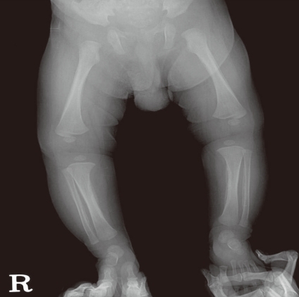
No organism was identified in any culture specimen, such as blood, urine or CSF. Although radiographic examination of the infant's long bones showed diaphyseal periostitis (Fig. 2), no abnormality was detected on brain magnetic resonance imaging. And hearing test was normal. After we diagnosed his illness as congenital syphilis, his parents were tested for syphilis and the mother's VDRL was found to be reactive with 1:4 dilutions and the father's VDRL was found to be reactive with a 1:1 dilution. Nevertheless this result, his mother had no specific symptoms. Based on these findings, the mother's VDRL results during prenatal care were erroneous.
We treated the patient with procaine penicillin G for 14 days. His skin eruption was resolved within several days. After 4 days of penicillin, results of serology test returned to normal with platelet count of 235,000/µL, WBC count of 13,670/mm3. During two weeks of hospitalization, the anemia slowly improved. Hemoglobin was increased to 10.0 g/dL with hematocrit 31.4%. Also, total protein was elevated slowly to 5.6 g/dL with albumin 4 g/dL. His parents were also treated with benzathine penicillin G. The patient was 5 month olds at the last visit and his physical examination was normal, including neurologic examination. In addition, the baby showed a reduction in seroreactivity from 1:32 to 1:8 dilutions.
Discussion
This 3-month-old infant presented only with skin eruption, which led to difficulty in diagnosis. Several physicians could not establish what his illness was, and his symptoms were misinterpreted as adverse reactions caused by DTaP vaccination. In addition, the maternal self report that she was VDRL-negative caused a further delay in diagnosis of the patient. Diagnosis of early congenital syphilis is difficult because more than half of the infants are asymptomatic, and signs in symptomatic infants may be subtle and nonspecific4). We found a report of an infant with a skin rash whose mother had adequate prenatal care, similar to our patient2). If diagnosis is missed, death may result, despite the fact that syphilis is a very easy and inexpensive disease to treat5).
Congenital syphilis occurs when T. pallidum crosses the placenta from the mother to the fetus during pregnancy or by contact with an infectious lesion during birth6). Manifestations of congenital syphilis are divided into early and late signs based on the first 2 years of life. Mucocutaneous involvement is present in as many as 70 percent of infants and may be apparent at birth or develop during the first few weeks of life6). Cutaneous findings of early congenital syphilis is classically a vesiculobullous or maculopapular rash on the palms and soles and may be associated with desquamation7). Other types of rashes such as erythema multiforme have also been reported7,8). In addition, symptoms of early congenital syphilis include fever, failure to thrive, hepatosplenomegaly, lymphadenopathy, osteochondritis, pneumonitis, and rhinitis6). Leukocytosis, Coombs-negative hemolytic anemia, thrombocytopenia, hypoproteinemia, hypoalbuminemia, hyperbilirubinemia, and elevated liver enzyme levels may be present6,9). Because these laboratory findings are difficult to identify upon physical examination, a high index of suspicion is necessary to make the right diagnosis early. Although acral dermatitis, vitamin or nutrient deficiency, and hand eczema might mimic this disorder, skin rash of early congenital syphilis is relatively recalcitrant to classical eczema treatment, which might be differential diagnostic point10,11). Characteristic mucocutaneous rash, presenting with erythematous maculopapular or bullous lesions, followed by desquamation involving hands and feet, are common in congenital syphilis12).
For definitive diagnosis, the Centers for Disease Control (CDC) recommends identification of syphilis in the mother; lack of evidence of adequate maternal treatment; presence of clinical, laboratory or radiological evidence of syphilis in the infant; and comparison of maternal and infant non-treponemal serologic titers using the same test and preferably the same laboratory, as was conducted in this case13). In addition, The CDC recommends serologic VDRL testing of pregnant women during the first prenatal visit and additional serologic testing and evaluation of sexual history at 28 weeks of gestation and soon after delivery in communities in which there is a high risk of congenital syphilis13).
Congenital syphilis is a preventable and treatable disease if physicians are aware of its diverse clinical symptoms. Therefore, clinical suspicion and formal confirmation of antenatal screening results as well as a detailed maternal history provide important clues for the diagnosis of congenital syphilis.

 PDF Links
 PubReader
 PubMed
 Download Citation


