

Introduction
The first severe case of H1N1 influenza-related pneumonia in a young adult was reported in April 20091), and the 2009 H1N1 influenza A virus spread rapidly thereafter. The World Health Organization (WHO) declared a pandemic in June 2009. By February 2010, at least 15,921 fatal cases as confirmed by laboratory tests were reported by the WHO2). In South Korea, the first fatal case was reported in August 2009, and 196 intensive care unit (ICU) patients were reported by December 7, 20093). Some patients with H1N1 influenza virus infection experienced rapidly progressing respiratory symptoms and ultimately required respiratory support, including mechanical ventilation, nitric oxide inhalation, and/or extracorporeal membrane oxygenation (ECMO). In Canada, over 80% of ICU patients required mechanical ventilation and of those patients 17 to 40% died4). It was recently found that 64.6% of H1N1 ICU patients in Australia and New Zealand needed mechanical ventilation during the winter of 2009 and that 11.6% of those patients were subsquently treated with ECMO5,6). In this report, we present a child with acute respiratory distress syndrome (ARDS) due to a 2009 H1N1 influenza virus infection complicated by necrotizing pneumonia who was successfully rescued with ECMO.
Case report
A 3-year-old girl was referred to the Samsung Medical Center, Seoul, Korea emergency department due to fever, respiratory distress, and decreased mental status during the peak influenza pandemic period in October 2009. She had been healthy until she developed fever and cough 7 days prior to hospitalization. Two days after her initial symptoms, she was seen by a general practitioner and was started on oseltamivir based on a positive rapid antigen test result. However, her condition deteriorated and she experienced difficult breathing and sleepiness despite antiviral treatment. She was taken to a general hospital near her home and was eventually referred to our center due to impending respiratory failure and decreased mental status. She did not have any significant past medical or family history, and she had not been vaccinated against 2009 H1N1 influenza or seasonal influenza.
On arrival, the patient's blood pressure was 117/67 mmHg, heart rate 181 beats/min, respiratory rate 40/min, and body temperature 37.5℃. Percutaneous oxygen saturation was 91% on 7 liters/min of oxygen via face mask. On physical examination, chest wall retraction was observed and left lung sounds were decreased. The patient did not have any abnormal focal neurological signs although she appeared drowsy. She was admitted to the pediatric intensive care unit and intubated immediately.
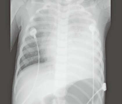
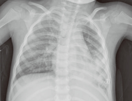
Her initial blood gas analysis showed pH 7.193, PaCO2 53.9 mmHg, PaO2 30.3 mmHg, bicarbonate 20.3 mmoL/L, and base excess -7.9 on 7 liters/min of oxygen via face mask. A complete blood count showed pancytopenia (white blood cell [WBC] count 710/µL, absolute neutrophil count 210/µL, hemoglobin 10.6 g/dL, platelets 81,000/µL) and her C-reactive protein was markedly elevated up to 37.18 mg/dL. Her immunoglobulin G level was decreased to 399 mg/dL. The patient was placed on high dose oseltamivir (60 mg/dose, body weight 13 kg) and intravenous immunoglobulin G was also given as 500 mg/kg/day for 2 days. The 2009 H1N1 influenza virus was detected by reverse transcriptase-polymerase chain reaction assay in both a respiratory specimen and spinal fluid. Her initial chest radiograph showed diffuse haziness in the entire left lung field and the right upper lung field (Fig. 1). Thoracentesis was performed and 100 mL of turbid chocolate-colored fluid was drained from the left lung (pleural fluid analysis results: red blood cell >1,000/µL, WBC >1,000/µL, polymorphonuclear neutrophil 93%, pH 7.4, glucose 11 mg/dL, protein 3,564.6 mg/dL). A culture of the pleural fluid did not grow any organisms.
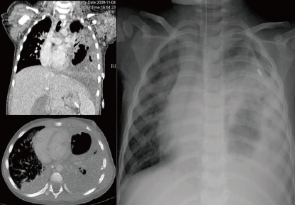
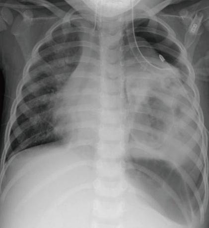
On hospital day 3, a chest tube was inserted into the left pleural space due to increased pleural effusion causing tracheal deviation. On hospital day 6, chest computed tomography showed necrotizing pneumonia with a significant left pleural effusion (Fig. 2). She also had persistent high fever (39.1℃) and a striking leukocytosis (WBC count 24,250/µL, absolute neutrophil count 19,640/µL) despite broad spectrum antibiotics (cefotaxime and vancomycin). On hospital day 7, pneumothorax developed in the left lung (Fig. 3) and the chest tube was replaced by one with a bigger sized lumen. As pneumothorax was waxed-and-waned on hospital day 8, more ventilator support (volume control mode, FiO2 1.0, positive end-expiratory pressure [PEEP] 5 cmH2O, TV 90 mL) was needed to maintain percutaneous oxygen saturation level near 90%. Peak inspiratory pressure was recorded at 23 to 34 cmH2O depending on the relief of pneumothorax. Due to her poor response to antibiotic treatment, cefotaxime was changed to meropenem. On hospital day 9, percutaneous oxygen saturation level was dropped to 77% due to progressive respiratory failure with a persistent purulent pleural effusion and a recurrent, massive pneumothorax (Fig. 4). To rescue the patient from progressing respiratory failure without raising positive airway pressure, which might aggravate pneumothorax, venovenous ECMO support was initiated.
Of note, multidrug resistant Acinetobacter baumannii started to grow from both blood and tracheal aspirate cultures on hospital day 10. Follow-up cultures persistently grew A. baumannii and Stenotrophomonas maltophilia from both tracheal aspirates and pleural fluid. Blood cultures became negative 5 days after ECMO discontinuation (bacteremia persisted from day 10 to day 18). The tracheal aspirate cultures were positive from hospital days 10 to 34, and pleural fluid cultures were positive from hospital days 11 to 41. The total duration of ECMO support was 6 days, and successful weaning was achieved without cardiovascular complications. At the time of ventilator weaning, blood gas analysis showed pH 7.371, PaCO2 64.0 mmHg, PaO2 111.9 mmHg, bicarbonate 36.3 mmoL/L, and base excess 9.2 with conventional mechanical ventilation. Various combinations of antibiotics were used including isepamicin, minocyclin, rifampicin, ceftazidime, TMP-SMX, and colistin in an attempt to control persistent infections due to multidrug resistant A. baumannii and S. maltophilia. Total duration of oseltamivir medication was 23 days (high dose for 18 days and usual dose for 5 days).
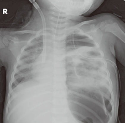
The patient was weaned from mechanical ventilation on hospital day 37, and all antimicrobials were discontinued on day 57. On day 63, the left chest tube was removed and she was discharged from the hospital alert and spontaneously breathing without difficulty in room air (Fig. 5). At the time this manuscript was submitted, the patient had no respiratory symptoms and a chest radiograph was markedly improved.
Discussion
About 1-10% of patients with clinical illness due to the H1N1 influenza virus infection were hospitalized, and the overall case fatality ratio has been estimated to be <0.5%7). Patients with dyspnea, tachypnea, evidence of hypoxemia, and pulmonary infiltrates on chest radiograph should be hospitalized8), and most deaths have been related to respiratory failure resulting from severe pneumonia with multifocal infiltrates and ARDS9,10). Mechanical ventilation is usually necessary to support gas exchange in patients with ARDS, although its use can be associated with lung injury. Despite attempts to optimize ventilator settings, hypoxemia or hypercapnia refractory to conventional mechanical ventilation may persist in a small subset of patients, and these patients may be treated with ECMO11). Furthermore, The Korean Society of Critical Care Medicine recommended early ECMO application if ARDS were not be maintained appropriately12). Recent studies have also shown significant improvement in the survival of patients transferred to a specialist center for ECMO treatment compared to conventional management13). This patient had recurrent, massive pneumothorax and pleural effusion, we were concerned that ventilation with high PEEP might aggravate lung parenchymal destruction and air leak and decided ECMO support in this patient.
In general, venovenous ECMO is used for isolated pulmonary support in cases of reversible pulmonary failure14). In older children, the most common diagnoses for ECMO support are viral pneumonia, bacterial pneumonia, ARDS, and aspiration pneumonia15). On the other hand, active irreversible respiratory injury is not an adequate indication for ECMO support. Although some complications associated with ECMO such as coagulopathy, thromboembolism could happen, the worldwide pandemic H1N1 influenza virus in 2009 caused many serious cases of ARDS and some of those patients were successfully treated with short term ECMO support5).
H1N1 influenza virus infection caused severe lung parenchymal destruction in certain patients, and these patients often do not respond to conventional respiratory care. ECMO support is recommended as the alternative treatment modality, and it is occasionally the only rescue method in pediatric patients.
The patient in this report had serious lung parenchymal injury, was septic, and had an uncertain neurological status. The severe lung parenchymal destruction (initially due to influenza virus and then secondary bacterial infection) with massive pneumothorax was responsible for the subsequent acute respiratory distress syndrome (ARDS) which was resistant to conventional mechanical ventilation. ECMO and aggressive antimicrobial treatment were used simultaneously to allow for oxygenation and to control the bacterial coinfection.
Streptococcus pneumoniae, Streptococcus pyogenes, Staphylococcus aureus, Streptococcus mitis, and Haemophilus influenza have all been identified as co-pathogens among the fatal H1N1 viral infection cases described in the US16). However, our patient developed secondary bacterial pneumonia and bacteremia due to a nosocomial infection; A. baumannii is a well-known gram-negative pathogen responsible for healthcare-associated infection in the ICU17).
In conclusion, we report a 3-year-old female patient with an acute H1N1 influenza virus infection and necrotizing pneumonia rescued with ECMO. This is the first reported successful pediatric ECMO rescue case in South Korea during the pandemic period. We will continue to follow up our patient's growth, lung function, and neurological development.
 PDF Links
 PubReader
 PubMed
 Download Citation


