 About
About Browse articles
 For contributors
All issues > Volume 69(5); 2026
Classification of neurocognitive impairment in pediatric drug-resistant focal epilepsy by quantifying seizure-affected brain network abnormalities in clinical diffusion-weighted imaging connectome
-
Jeong-Won Jeong, PhD1,2,3,4
 , Min-Hee Lee, PhD1,2, Yoon Ho Hwang, PhD1,2, Michael Behen, PhD1,3, Aimee Luat, MD3,5, Csaba Juhász, MD, PhD1,2,3,4, Eishi Asano, MD, PhD1,2,3,4,5
- Corresponding author: Jeong-Won Jeong, PhD. Professor of Pediatrics, Neurology, and Translational Neuroscience Program, Wayne State University School of Medicine, Detroit, MI USA Email: jjeong@med.wayne.edu
- Received December 18, 2025 Revised January 30, 2026 Accepted February 5, 2026
- Abstract
-
- Background
- Background
- Diverse factors including seizure onset age, seizure frequency, epilepsy duration, total number of antiseizure medications trialed are considered as seizures-related neurocognitive loads in children with drug-resistant focal epilepsy (DRE). However, their associations with the structural integrity of neurocognitive networks remain largely unknown.
- Purpose
- Purpose
- This study investigates a novel diffusion-weighted imaging (DWI) connectome methodology that can extract seizure-associated structural abnormality biomarkers from clinical DWI tractography, use them to classify neurocognitive impairments prior to surgery, and unveil the relationship between epilepsy-related factors and neurocognitive impairments.
- Methods
- Methods
- Thirty-three DRE children (age: 11.8±3.3 years, 17 boys) and 29 age-matched healthy controls were enrolled to create seizure-affected networks whose edges connect epileptogenic regions to key brain regions of 6 neurocognitive networks. The deviations of local efficiency values were averaged across the seizure-affected brain regions and used as new imaging-based biomarkers quantifying the degrees of seizure-associated structural abnormalities accumulated on individual neurocognitive networks and classifying the neurocognitive impairments along with the epilepsy-related factors.
- Results
- Results
- Effect sizes of the proposed biomarkers for differentiating DRE from healthy controls were consistently very large across various subgroups defined by lesion types, lobar locations of epileptogenic foci, seizure frequency categories, and seizure types (i.e., Cohen d value >1.8). Compared with the epilepsy-related factors, the proposed biomarkers demonstrated superior classification accuracy for identifying neurocognitive impairments in general, verbal, and nonverbal domains. When combined with the epilepsy-related factors, the classification performance further improved, achieving an accuracy range of 90%–98% in the independent test patients. The subsequent association analysis using the proposed biomarkers as seizure-associated structural abnormality indicators demonstrated that the inclusion of such imaging indicators significantly enhances the strength of associations between epilepsy factors and neurocognitive impairments.
- Conclusion
- Conclusion
- These findings offer strong potential for objectively identifying neurocognitive impairments in DRE children, supporting early, data-driven decisions for personalized interventions to mitigate long-term effects.
- Introduction
- Introduction
Despite the availability of over 20 antiseizure medications (ASMs), approximately one-third of pediatric patients with epilepsy experience medically refractory seizures - commonly referred to as drug-resistant focal epilepsy (DRE) [1]. This condition leads to a significantly elevated risk of diverse neurocognitive impairments, including intelligence quotient (IQ) and language concerns [2,3]. Throughout the early stages of brain development, the brain undergoes rapid maturation of white matter tracts and large-scale neural network integration [4,5]. These developmental processes are highly susceptible to disruption from drug-resistant seizures, which can interfere with normal neurodevelopmental trajectories [6,7]. Consequently, pediatric DRE has an urgent need for more effective management and earlier intervention, which can mitigate the significant risk of long-term neurocognitive declines.Recent advances in diffusion-weighted imaging connectome (DWIC) analysis advances more precise, noninvasive imaging of white matter organization and brain network topology in children with DRE [8,9]. Our prior DWIC works [10,11] also found significantly reduced structural connectivity strength, reduced local efficiency of neighboring connections, and abnormal structural hub configuration across whole-brain regions, suggesting that DRE with high treatment tolerance may be associated with widespread network-level disorganization of white matter connectivity. However, the specific impact of seizure load [12,13], “the cumulative burden of seizure activity,” on the organization of specific neurocognitive networks remains poorly understood. For instance, its causal association with epilepsy-related factors (e.g., seizure onset age, seizure frequency, epilepsy duration, total number of ASM trialed) remains uncertain. Also, the extent to which neurocognitive dysfunction directly results from abnormal white matter organization within seizure-affected network remains unclear, leaving a critical gap in understanding how seizure-associated structural abnormalities interferes with neurocognitive trajectories in individual children with DRE.This study introduces a novel DWIC-based preoperative evaluation tool for children with DRE that utilizes their clinical DWIC data to: (1) construct a seizure-affected network with pairwise white matter tract pathways connecting seizure onset zones (SOZs) to other brain regions of interest, called “nodes,” that are involved in 6 neurocognitive domains – full scale IQ, verbal IQ, nonverbal IQ, core language, expressive language, and receptive language; (2) use individual children’s seizure-affected network to extract an imaging-based seizure-associated structural abnormality biomarker for each of the 6 neurocognitive domains by averaging deviations of local efficiency values in SOZ-connected nodes of each neurocognitive domain; (3) use a multiple layer perceptron (MLP) neural network with the extracted seizure-associated structural abnormality biomarkers to classify neurocognitive impairments in general, verbal, and nonverbal domains; and (4) use partial least square structural equation modeling (PL-SEM) [14,15] to uncover the underlying relationship between the extracted seizure-associated structural abnormality biomarkers and varying degrees of neurocognitive impairment severity, as determined by preoperative neuropsychological evaluations.The working hypothesis of this study is that DRE without effective therapeutic intervention (e.g., ASM, surgery) leads to a higher seizure load which contributes to more pronounced abnormality in the seizure-associated structural abnormality biomarker highlighted by more disruption of axonal connectivity in brain nodes of 6 different neurocognitive networks connected to SOZs and results in more pronounced impairments in general, verbal, and nonverbal cognitive domains. The present study anticipates that the relationship between the seizure-associated structural abnormality biomarker and neurocognitive impairments may offer neurological evidence elucidating the impact of seizure load on the risk of various neurocognitive impairments. Based on this premise, the findings of this study would support the significance of early surgical intervention in mitigating the risk of neurocognitive deficits by reducing seizure load on the developing neurocognitive networks. Therefore, this new biomarker could help meet an important need in the field by improving the accuracy of individual seizure load assessments and aiding in the timely identification of patients who would benefit from surgery, thereby reducing the risk of neurocognitive decline.
- Methods
- Methods
- 1. Subjects
- 1. Subjects
This study enrolled 33 children clinically diagnosed with DRE (age: 11.8±3.3 years, 17 boys, Table 1) who underwent 2-stage resective surgeries at the Children’s Hospital of Michigan, Detroit, between 2009 and 2024. DRE was defined according to the International League Against Epilepsy criteria as failure of adequate trials of 2 or more appropriate ASMs to achieve sustained seizure freedom [16]. These patients were selected by applying the following inclusion criteria: (1) age: 2.5–19 years; (2) a history of DRE scheduled for intracranial electroencephalogram (iEEG) recording as a part of their pre-surgical evaluation; and (3) preoperative neuropsychological evaluation and clinical magnetic resonance imaging (MRI) acquisition including diffusion-weighted imaging (DWI) tractography scan were successfully completed. Exclusion criteria consisted of the following: (1) significant brain malformations (such as large perisylvian polymicrogyria or hemimegalencephaly), as excessive anatomical disruptions preclude accurate coregistration with a standard anatomical template; (2) history of previous neurological surgery; and (3) autism spectrum disorder based on clinical and neuropsychological evaluation.Twenty-nine healthy children (age: 11.6±3.3 years, 14 boys) were also enrolled as a control group, matched to the DRE children by age, sex, and MRI protocol, and used to calculate a normative range of the seizure-associated structural abnormality biomarker for evaluating age-matched DRE patient’s deviation of the seizure-associated structural abnormality biomarker. Our DRE study cohort (n=33) was divided into 2 separate cohort sets, (1) development set (n=20) to train and validate the performance of the proposed MLP model and (2) independent test set (n=13) to validate the reproducibility of the trained MLP model on an independent study cohort that was not included in the model development. The 2 sets were statistically matched for age-sex-seizure-affected hemisphere-etiology-epilepsy duration using standardized mean differences (SMDs) [17] to ensure comparability (i.e., SMD values of age, sex, affected hemisphere, etiology, and epilepsy duration were 0.004, 0.077, 0.023, 0.094, and 0.097, respectively, which meets the threshold of less than 0.1 indicating good similarity). This study was approved by an Institutional Review Board (IRB) of Wayne State University (IRB number 111014MP2F), and written informed consent or a waiver of consent was obtained from the patients and/or their guardians of the patients.- 2. Neurocognitive evaluation
- 2. Neurocognitive evaluation
Age-appropriate measures of language processing were performed using Comprehensive Evaluation of Language Fundamentals Edition (CELF) including the CELF-Preschool Edition and CELF-4 that yield standardized indices of core, receptive, and expressive language processing from individual DRE children. Global, verbal, and nonverbal intellectual functioning were also assessed using the age-appropriate versions of the Wechsler series: the Wechsler Preschool and Primary Scale of Intelligence for children 2.6 through 6.0 years of age, the Wechsler Intelligence Scale for Children for children 6 years through 16 years of age, and the Wechsler Adult Intelligence Scale for individuals older than 16 years of age. Time interval between MRI and neuropsychological assessment was 2.4±4.6 months.- 3. Subgrouping of neurocognitive impairment severity
- 3. Subgrouping of neurocognitive impairment severity
According to the Wechsler classification scheme, individual’s performance in class I: general cognitive ability, class II: verbal ability, and class III: nonverbal ability was then categorized into, score (1) high average (standard score [SS] >110); score (2) average (SS=90–110); score (3) low average (SS=80–89); score (4) borderline (SS=70–79); score (5) mildly impaired (SS=60–69); score (6) severely impaired (SS<60). The 6 subgroups in class I/II/III—high average (n=3/3/2), average (n=8/6/11), low average (n=6/6/7), borderline (n=8/5/2), mildly impaired (n=3/5/2), severely impaired (n=3/6/4)—were utilized to investigate the effectiveness of the proposed seizure-associated structural abnormality biomarker for improving the PL-SEM based association model.- 4. MRI acquisition
- 4. MRI acquisition
All participants underwent clinical brain MRI scanning using a 3T GE SIGNA scanner (GE Healthcare, USA) equipped with an 8-channel head coil. DWI was acquired with the following parameters: repetition time (TR)=12,500 msec, echo time (TE)=86.4 msec, flip angle=90°, slice thickness=3 mm, 55 isotropic gradient directions at b=1,000 s/mm2, and single b=0 s/mm² image acquisition. T1-weighted images were obtained using a 3-dimentional fast spoiled gradient echo sequence with TR=6.1 ms, TE=2.1 ms, flip angle=12°, and slice thickness=1.2 mm.- 5. Construction of neurocognitive networks from whole-brain DWIC
- 5. Construction of neurocognitive networks from whole-brain DWIC
Diverse DWI artifacts including head motion, noise, physiological artifacts, susceptibility-induced distortion, B1 field inhomogeneity, and eddy current-induced distortion were corrected using the FSL (FMRIB [Oxford Centre for Functional MRI of the Brain] Software Library) tools [18,19] and a deep learning-based distortion correction approach [20]. We utilized MRtrix package (http://www.mrtrix.org/) to generate 50 million tracts by applying SIFT1 reconstruction to 100 million iFOD2-ACT whole-brain tracts [21]. Our deep convolution neural network-based tract classification [22] removed false-positive tracts (e.g., wiggly tracked fiber, broken fiber) from the iFOD2-ACT whole-brain tractography of individual participants. Finally, the resulting true positive whole-brain tractography was used to construct a whole backbone DWIC graph, G=(W,S), where Wi represents a set of 724 brain nodes, and each element Si,j represents an edge (or connectivity strength) quantifying the average fractional anisotropy (FA) value of the fiber tracts connecting Wi and Wj. To define a set of 724 brain nodes, Wi=1-724, this study combines 690 nodes of a Yale brain atlas [23] with 8 subcortical and 26 cerebellar nodes of an automated anatomical labeling atlas [24].- 6. Construction of seizure-affected network for seizure-associated structural abnormality biomarker
- 6. Construction of seizure-affected network for seizure-associated structural abnormality biomarker
Six neurocognitive networks, Nk=1-6(m,n), were constructed using our previous psychometry-driven diffusion tractography network analysis [11] by gathering all elements of S(i,j) of which values are significantly correlated with the intersubject scores of full scale IQ (N1), verbal IQ (N2), nonverbal IQ (N3), core language (N4), expressive language (N5), and receptive language scores (N6). In each Nk of a given patient we identified all brain nodes directly connected to iEEG-defined SOZ sites and averaged their z scores of local efficiency [25] as a new seizure-associated structural abnormality biomarker:- 7. Data analysis and statistics
- 7. Data analysis and statistics
Three multilayer perceptron (MLP) utilized ek=1-6 as their inputs to classify the binary outputs: presence of neurocognitive impairment (i.e., 0 and 1 for no and yes corresponding to the ground truth score of general, verbal, and nonverbal ability less than or equal to and greater than 3, respectively). Four epilepsy-related factors ck=1-4 including seizure onset age (c1), seizure frequency (c2), epilepsy duration (c3) and total number of ASM trialed (c4) were used as non-imaging variables for the comparison. The MLP architecture consisted of 2 hidden layers with 64 and 32 units, respectively. Input features were standardized prior to training, and a regularization parameter of 0.01 was applied to prevent overfitting. Network weights were initialized using the He method.Model evaluation was performed using leave-one-subject-out cross-validation (LOSOCV), in which the data from one subject was reserved for validation, while the data from the remaining subjects (i.e., n=19) in the development set were used for training in each fold. To address overfitting with small sample sizes, data augmentation was performed on both ek=1-6 and ck=1-4 using the synthetic minority oversampling technique [26]. For each cross-validation training split, the 4 nearest neighbors were used to generate synthetic instances, and 100 additional samples were generated.The generalizability of the trained MLP was evaluated on an independent test set where ck=1-4 and ek=1-6 were obtained using the same procedure as applied to the development set. The overall classification performance for identifying neurocognitive impairments was evaluated using 3 metrics: (1) balanced accuracy (BA), (2) the area under the receiver operating characteristic curve [27], and (3) the area under the precision-recall curve [28]. For each of the 20 models trained through LOSOCV, the values of these metrics were computed and then averaged. To evaluate the extent of potential overfitting, we also performed a random label learning experiment by repeating both the training and testing procedures of the MLP with pseudo neurocognitive impairments that were randomly assigned to individual subjects in the neurocognitive outcomes: 0 or 1. It was assumed that if the BA values resulted from this experiment were less than 0.5, the impact of overfitting at the given sample size would be minimal.Finally, the present study utilized PL-SEM approach [14,15] to elucidate the strengths of associations by which an unmodifiable latent variable of epilepsy severity indicators (i.e., seizure onset age, seizure frequency) and a modifiable latent variable of treatment indicators (i.e., total number of ASM trialed, epilepsy duration at the time of epilepsy surgery) jointly influence a latent variable of neurocognitive impairment indicators (i.e., class I: general cognitive ability score, class II: verbal ability score, class III: nonverbal ability score), while investigating whether these associations are mediated by an imaging latent variable of seizure-affected network abnormality indicators (i.e., ek). Using the general workflow of PL-SEM based association inference, we examined how the unmodifiable variable interplays with the modifiable variable and the imaging variable prior to affecting neurocognitive impairment variable. In this context, we designed 2 pathway analyses to investigate whether the inclusion of the imaging variable enhances the robustness of associations among unmodifiable variables, modifiable variables, and neurocognitive impairment variables: (i) unmodifiable→modifiable→neurocognitive impairment and (ii) unmodifiable→modifiable→imaging→neurocognitive impairment.Age and sex were included as covariates to control for potential confounding effects. Path coefficient (β), determination coefficient (r2), and the effect size of path coefficients (f2) were used to evaluate both direction and strength of association effects. The significance of model parameters and indicator loadings across all paths was estimated using bootstrap resampling procedures with 1,000 samples to infer the following hypotheses, (1) in both (i) and (ii), the modifiable variable has a statistically significant relationship with the modifiable variable (i.e., the P value of path coefficient <0.05), underscoring the importance of early therapeutic treatment indicators to mitigate the impact of the unmodifiable risk indicators on neurocognitive impairment; (2) both r2 and f2 values of the imaging variable in (ii) are greater than those of the modifiable variable in (i), indicating that integrating the imaging variable yields more robust and stable association between the unmodifiable variable and neurocognitive impairment.
- Results
- Results
- 1. Extraction of seizure-affected network biomarkers
- 1. Extraction of seizure-affected network biomarkers
Fig. 1 presents representative 3-dimentional visualizations of seizure-affected network identified in patients with left temporal lobe epilepsy and right temporal lobe epilepsy, respectively. In each patient, 6 neurocognitive networks, Nk=1-6(m,n) were constructed by our previous psychometry-driven tractography network analysis [25] at the P value of 0.01, after corrected for age and sex. A total of 277, 227, 183, 328, 362, and 223 nodes were identified as hub regions of N1(m,n), N2(m,n), N3(m,n), N4(m,n), N5(m,n), and N6(m,n), respectively. The z scores of their local efficiency values were then averaged to calculate the seizure-affected network biomarker, ek, for each patient. Notably, unilateral temporal SOZ sites exert a stronger influence on brain nodes located in the ipsilateral temporal and occipital regions within each Nk, in both cases.- 2. Classification of neurocognitive impairment using the seizure-affected network biomarkers
- 2. Classification of neurocognitive impairment using the seizure-affected network biomarkers
Fig. 2A shows significant separability of the proposed seizure-affected network biomarker, ek=1-6, to differentiate DRE children from age-matched healthy controls, evidencing a very large effect size of seizure-caused local efficiency reduction accumulated in neighboring connections of the given kth neurocognitive network Nk. Cohen d values of ek=1-6 between control and DRE groups were 1.87±0.08 and 1.93±0.06 in the development and test set, respectively. They did not differ at the P value of 0.15, suggesting the reproducible separability across 2 study sets. In addition, it was found that DRE originating from SOZ sites resulted in greater white matter abnormalities at the seizure-affected network nodes structurally connected to the SOZ sites, compared to those nodes not connected across Nk=1-6 (Fig. 2B). Furthermore, the effect size of ek=1-6 was consistently very large across various subgroups defined by lesion types, lobar locations of SOZ sites, seizure frequency categories, and seizure types (Fig. 3), indicating strong discriminative power for distinguishing heterogeneous DRE populations from controls.Table 2 suggests that the proposed ek=1-6 can outperform ck=1-4 in classifying the presence of impairment in class I: general ability, class II: verbal ability, and class III: nonverbal ability (with an average improvement of up to 32% BA). The highest accuracy (0.90–0.98) was obtained when the presence of each impairment was classified from ck=1-4 and ek=1-6. Compared to the random label experiment, the true label experiment produced substantially higher performance metrics across the test dataset. This indicates that the MLP was able to effectively learn detailed features of ck=1-4 and ek=1-6, enabling accurate classification of the presence of neurocognitive impairments with minimal overfitting, despite the limited sample size of the development set (n=20). The result of this comparison suggests a high potential of the proposed biomarkers, ck=1-4 and ek=1-6 to facilitate time-consuming seizure load assessments in clinical cases.- 3. Assessment of the association between seizure-associated structural abnormality biomarkers and neurocognitive impairments
- 3. Assessment of the association between seizure-associated structural abnormality biomarkers and neurocognitive impairments
Fig. 4 presents the association mechanisms among multiple latent variables of 2 directional pathways that were designed to study the effect of the imaging seizure-affected network abnormality on the neurocognitive impairment, (i) unmodifiable epilepsy severity→modifiable treatment→neurocognitive impairment and (ii) unmodifiable epilepsy severity→modifiable treatment→imaging seizure-affected network abnormality→neurocognitive impairment. In both pathways of (i)/(ii), path coefficient and effect size of the start path: unmodifiable epilepsy severity→modifiable treatment were statistically significant and sufficiently large (P=0.046/0.025, f2=0.71/0.79, respectively) at our sample size (n=33), suggesting the existence of the strong relationship between epilepsy severity and intervention. Higher path coefficient value and effect size were found in the end path: imaging seizure-affected network abnormality→neurocognitive impairment of pathway (ii) (β=0.63, P<0.001, f2=0.65), compared with those in the end path: modifiable treatment→neurocognitive impairment of pathway (i) (β=0.52, P=0.089, f2=0.37), indicating that the inclusion of the imaging variable further enhances the relationship between the modifiable variable and the neurocognitive impairment variable. The r2 values of individual latent variables were much higher in pathway (ii); this suggests that the inclusion of the proposed seizure-affected network-based imaging variable improves overall accuracy to classify the neurocognitive impairment from individual indicators used in the association model.
- Discussion
- Discussion
The present study introduced DWIC-based seizure-affected network biomarkers to characterize seizure-related structural network abnormalities in children with DRE. We found that SOZs were associated with altered multiple neurocognitive networks within temporal and occipital regions of the same hemisphere. The proposed biomarkers showed robust and reproducible differentiation between pediatric DRE patients and healthy controls, with consistently large effect sizes across independent datasets and heterogeneous clinical subgroups. Furthermore, these biomarkers demonstrated improved performance in classifying neurocognitive impairments compared with conventional epilepsy-related factors and were strongly associated with cognitive outcomes in multivariate pathway models. This suggests that seizure-related network abnormalities may partially mediate the relationship between a modifiable treatment-related latent variable and neurocognitive impairment in pediatric DRE. Our proposed biomarkers are designed to capture the cumulative network-level impact of seizure activity on connected brain networks, rather than direct measures of clinical seizure burden. Clinically, they are therefore conceptually distinct from conventional seizure burden metrics and provide complementary information regarding seizure-related network vulnerability and its potential contribution to neurocognitive impairment.In the past, numerous studies [29-31] have emphasized the impact of epilepsy-related factors, particularly seizure frequency and ASM burden, on neurocognitive development. The adverse effects of frequent seizures on neuropsychological functioning have been well documented in pediatric populations across various epilepsy syndromes, including generalized symptomatic epilepsy, idiopathic generalized epilepsy, temporal lobe epilepsy, and frontal lobe epilepsy. In particular, children with frontal lobe epilepsy [32] present with a wide range of neurocognitive impairments. Among the epilepsy-related variables, poor seizure control and the use of ASM polytherapy are most consistently associated with unfavorable neurocognitive outcomes. These associations suggest that epileptic discharges may interfere with the functional integrity of critical cerebral regions involved in cognitive processing. Furthermore, children with ongoing seizures despite pharmacological treatment exhibit significantly poorer cognitive performance compared to those achieving seizure control with monotherapy [32]. This observation may point to an underlying macrostructural brain pathology contributing to both seizure persistence and cognitive dysfunction. Although preliminary, our data also supports that macrostructural brain pathology reflects an atypical reduction in white matter integrity underlying significant axonal loss affected by the accumulated seizure loads, that was supported by more decrease in axial diffusivity (e.g., Cohen d=1.03) rather than more increase in radial diffusivity (e.g., Cohen d=-0.40), that indicates demyelination or myeline damage.In line with previous findings, the present study demonstrates that DRE originating from the SOZ sites are associated with widespread white matter abnormalities across various brain regions, indicating that the impact of DRE may extend beyond the confines of the epileptogenic zone. Notably, the effects of DRE associated with seizure frequency, duration of epilepsy, and total number of ASM trialed demonstrate a significant correlation with the proposed overall seizure-associated structural abnormality biomarkers (Fig. 5), where canonical correlation analysis was applied to explore the relationships between 2 sets of variables, different combinations of 3 epilepsy factors, and 6 seizure-associated structural abnormality biomarkers. These significant associations could justify the neurological basis of the proposed biomarkers in accurately quantifying the cumulative impact of seizure burden on individual neurocognitive impairments.The clinical translation of the proposed seizure-associated structural abnormality biomarkers has the potential to significantly improve epilepsy care by offering a reliable, objective, and comprehensive picture of how DRE impacts the brain, supporting better diagnosis, monitoring, and treatment strategies to prevent or mitigate long-term neurocognitive impairments. According to a large single site cohort study [29], prior to undergoing pediatric epilepsy surgery, 85% of patients exhibited neuropsychological impairments in at least one cognitive domain. Following surgery, the proportion of impaired patients decreased substantially, with 21%–50% demonstrating a transition from impaired to unimpaired status, and 16%–42% showing individually significant improvements. These findings underscore the efficacy of epilepsy surgery in children and suggest that postoperative functional gains, observed one year after intervention, may be influenced by factors such as the extent of baseline neurological impairment, cognitive reserve, neuroplasticity, preservation of functional brain tissue, and the beneficial effects of seizure freedom and reduced ASM burden. We anticipate that the proposed seizure-associated structural abnormality biomarkers may have potential value as exploratory metrics for characterizing neurocognitive impairment risk across various clinical settings in relation to surgical intervention. Given that diffusion tractography has become a standard component of preoperative MRI protocols in most epilepsy centers, the integration of these biomarkers into future routine practice may be feasible and potentially informative.The present study has multiple limitations. First, the 6 neuropsychological measures of cognitive functions used for the construction of seizure-affected network were inherently intercorrelated, leading to considerable overlap in key brain regions across the proposed biomarkers, ek=1-6. Consequently, information redundancy may be present, potentially contributing to model overfitting in the observed directional associations. Second, advanced assessment tools for epilepsy severity such as pediatric epilepsy severity scale [33], Chalfont seizure severity scale [34], national hospital seizure severity scale [35] were not considered with the proposed seizure-associated structural abnormality biomarkers. Thus, the findings of this study should be carefully considered as a proof-of-concept supporting that the advanced DWIC analysis holds potential to offer objective, imaging-based seizure-associated structural abnormality markers that may help address the current shortcoming of seizure load assessment tools that are particularly challenging to evaluate in pediatric populations. Third, the limited sample size in this study was largely due to missing data related to the assessment of neurocognitive impairment. This constraint affected the reliability of the intervention variable in the PL-SEM analysis, as reflected by suboptimal Cronbach α (0.50) and composite reliability (0.50) values. Although the intervention variable demonstrated statistically significant and substantial path coefficients and effect sizes across both directional pathways, these findings require validation in a larger sample to achieve acceptable reliability thresholds (i.e., Cronbach α and composite reliability >0.70) and to confirm reproducibility. Finally, the retrospective design of this study limited the ability to assess the efficacy of the proposed seizure-associated structural abnormality biomarkers using longitudinal data. As a result, the observed associations were derived from cross-sectional analyses, failing to investigate the actual causality. Thus, the reported findings should be interpreted as population-level association inferences rather than individual-level causal relationships. To establish the clinical utility and classification validity of these biomarkers, future research should incorporate prospective longitudinal causality study designs with repeated measurements at the individual level. In addition, although subgroup analyses by lesion type were performed, the present study cannot distinguish whether the proposed imaging biomarkers reflect seizure-related network propagation or the structural lesion impact. Lesion-related structural effects and seizure-related network abnormalities may interact, and causal separation is limited by the current cross-sectional study design. Further studies using prospective designs will be necessary to clarify the relative contributions of seizure-related and lesion-related mechanisms. In addition, the present study does not provide individual-level thresholds, prospective validation, or decision-impact analyses. Therefore, the proposed imaging biomarkers should be considered exploratory and hypothesis-generating, and extensive validation in independent and prospective cohorts will be required before clinical application.In conclusion, the present study demonstrates the effectiveness of our novel DWIC-based seizure-associated structural abnormality biomarkers that can objectively quantify cumulative network-level abnormalities associated with altered neurocognitive developmental trajectories in children with DRE. It was found that DRE originating from various SOZ sites propagate to many functional regions within various neurocognitive networks, directly leading to a significant reduction in local efficiency of adjacent white matter connections in individual neurocognitive networks. Consequently, this disruption was found to contribute to significant neurocognitive impairments through its significant associations with the proposed biomarkers.
- Footnotes
-
Conflicts of interest No potential conflict of interest relevant to this article was reported.
Funding The research reported in this article was supported by the National Institute of Health under Award Number NIH R01NS089659 (to JJ) and R01NS064033 (to EA). The content is solely the responsibility of the authors and does not necessarily represent the official views of the National Institutes of Health.
Author contribution Conceptualization: JJ, AL, CJ, EA; Data curation: JJ, MHL, YHH, MB, AL, CJ, EA; Formal analysis: JJ, MHL, YHH, MB, AL, CJ, EA; Funding acquisition: JJ, EA; Methodology: JJ, MHL; Project administration: JJ; Visualization: JJ, MHL; Writing - original draft: JJ; Writing - review & editing: JJ, MHL, YHH, MB, AL, CJ, EA
-
Fig. 1.
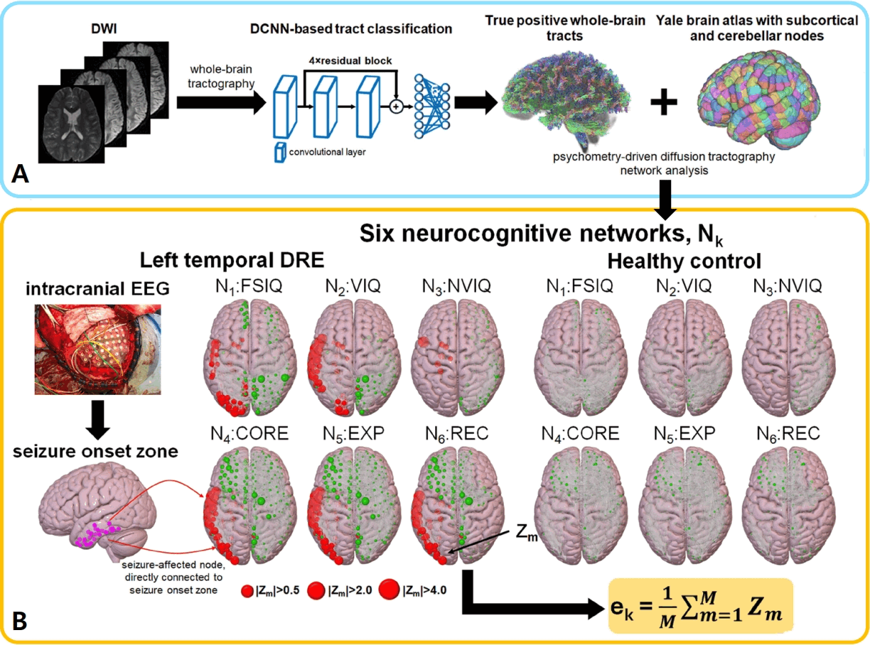
Fig. 2.
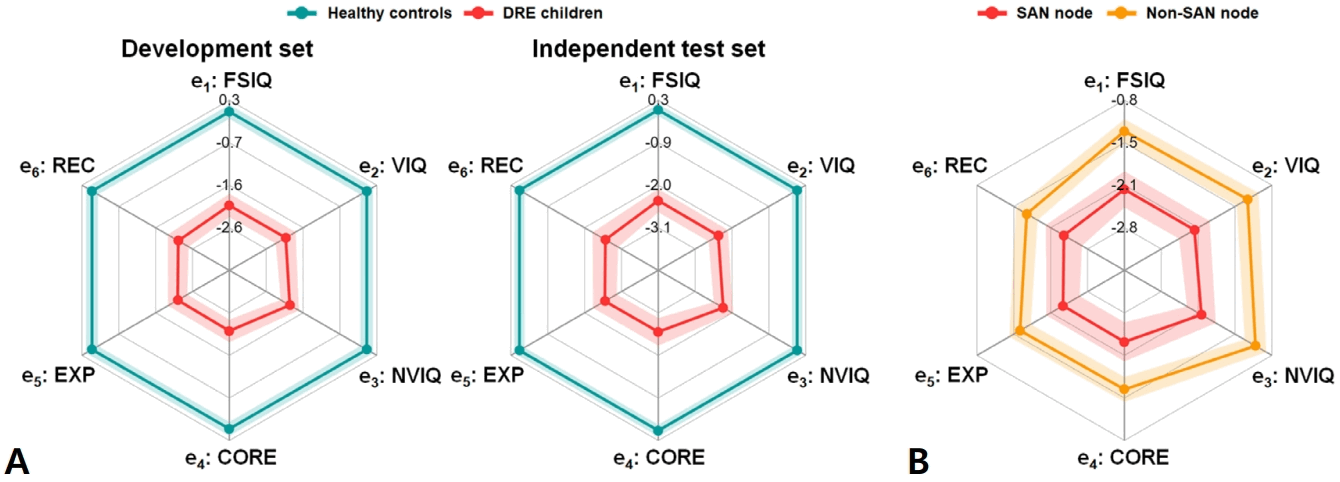
Fig. 3.
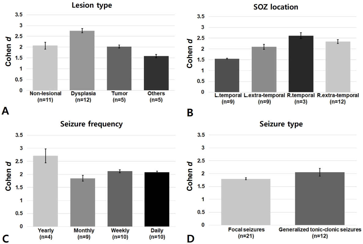
Fig. 4.
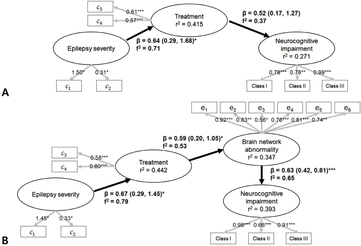
Fig. 5.
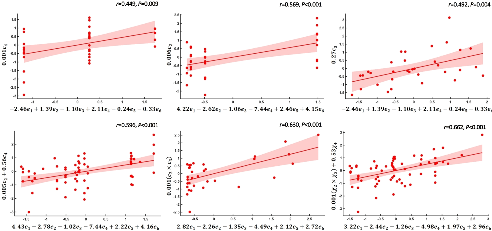
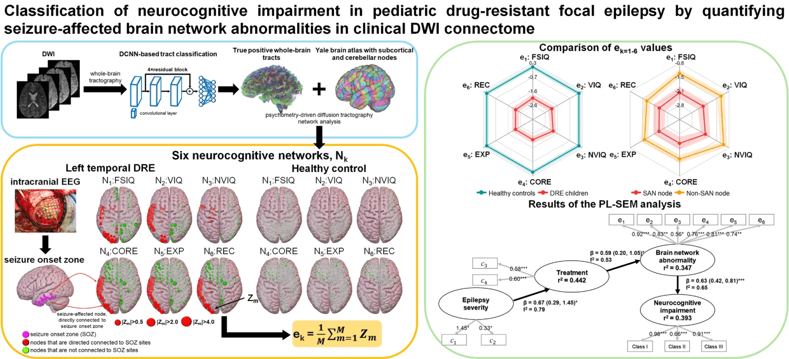
Table 1.
Table 2.
BA, balanced accuracy; AUROC, area under the receiver operating characteristic curve; AUPRC, area under the precision-recall curve.
Mean and standard deviation of each metric were evaluated using 20 LOSOCV-based multiple layer perceptron models with 3 different types of patient variables (ck=1-4, ek=1-6, ck=1-4, and ek=1-6).
- References
- 1. Löscher W, Potschka H, Sisodiya SM, Vezzani A. Drug Resistance in epilepsy: clinical impact, potential mechanisms, and new innovative treatment options. Pharmacol Rev 2020;72:606–38.
[Article] [PubMed] [PMC]2. Baumer FM, Cardon AL, Porter BE. Language Dysfunction in pediatric epilepsy. J Pediatr 2018;194:13–21.
[Article] [PubMed] [PMC]3. Warsi NM, Wong SM, Gorodetsky C, Suresh H, Arski ON, Ebden M, et al. Which is more deleterious to cognitive performance? Interictal epileptiform discharges vs anti-seizure medication. Epilepsia 2023;64:e75–81.
[Article] [PubMed]4. Hagmann P, Sporns O, Madan N, Cammoun L, Pienaar R, Wedeen VJ, et al. White matter maturation reshapes structural connectivity in the late developing human brain. Proc Natl Acad Sci U S A 2010;107:19067–72.
[Article] [PubMed] [PMC]5. Huang H, Shu N, Mishra V, Jeon T, Chalak L, Wang ZJ, et al. Development of human brain structural networks through infancy and childhood. Cereb Cortex 2015;25:1389–404.
[Article] [PubMed] [PMC]6. Stasenko A, Lin C, Bonilha L, Bernhardt BC, McDonald CR. Neurobehavioral and clinical comorbidities in epilepsy: the role of white matter network disruption. Neuroscientist 2024;30:105–31.
[Article] [PubMed] [PMC]7. Gilsoul M, Grisar T, Delgado-Escueta AV, de Nijs L, Lakaye B. Subtle brain developmental abnormalities in the pathogenesis of juvenile myoclonic epilepsy. Front Cell Neurosci 2019;13:433.
[Article] [PubMed] [PMC]8. Larivière S, Bernasconi A, Bernasconi N, Bernhardt BC. Connectome biomarkers of drug-resistant epilepsy. Epilepsia 2021;62:6–24.
[Article] [PubMed]9. Zhang Y, Jiang L, Zhang D, Wang L, Fei X, Liu X, et al. Thalamocortical structural connectivity abnormalities in drug-resistant generalized epilepsy: a diffusion tensor imaging study. Brain Res 2020;1727:146558.
[Article] [PubMed]10. Jeong JW, Lee MH, Behen M, Uda H, Gjolaj N, Luat A, et al. Quantitative phenotyping of verbal and non-verbal cognitive impairment using diffusion-weighted MRI connectome: preliminary study of the crowding effect in children with left hemispheric epilepsy. Epilepsy Behav 2024;160:110009.
[Article] [PubMed] [PMC]11. Lee MH, Banerjee S, Uda H, Carlson A, Dong M, Rothermel R, et al. Deep learning-based tract classification of preoperative dwi tractography advances the prediction of short-term postoperative language improvement in children with drug-resistant epilepsy. IEEE Trans Biomed Eng 2025;72:565–76.
[Article] [PubMed] [PMC]12. Kitazawa Y, Jin K, Kakisaka Y, Fujikawa M, Tanaka F, Nakasato N. Predictive factors of higher drug load for seizure freedom in idiopathic generalized epilepsy: comparison between juvenile myoclonic epilepsy and other types. Epilepsy Res 2018;144:20–4.
[Article] [PubMed]13. Hermann B, Seidenberg M, Bell B, Rutecki P, Sheth R, Ruggles K, et al. The neurodevelopmental impact of childhood-onset temporal lobe epilepsy on brain structure and function. Epilepsia 2002;43:1062–71.
[Article] [PubMed]14. Yeni K, Tulek Z, Simsek OF, Bebek N. Relationships between knowledge, attitudes, stigma, anxiety and depression, and quality of life in epilepsy: a structural equation modeling. Epilepsy Behav 2018;85:212–17.
[Article] [PubMed]15. Hair JF, Sarstedt M, Pieper TM, Ringle CM. The use of partial least squares structural equation modeling in strategic management research: a review of past practices and recommendations for future applications. Long Range Plann 2012;45:320–40.
[Article]16. Kwan P, Arzimanoglou A, Berg AT, Brodie MJ, Allen Hauser W, Mathern G, et al. Definition of drug resistant epilepsy: consensus proposal by the ad hoc Task Force of the ILAE Commission on Therapeutic Strategies. Epilepsia 2010;51:1069–77.
[Article] [PubMed] [PMC]17. Austin PC. Balance diagnostics for comparing the distribution of baseline covariates between treatment groups in propensity-score matched samples. Stat Med 2009;28:3083–107.
[Article] [PubMed] [PMC]18. Andersson JLR, Graham MS, Zsoldos E, Sotiropoulos SN. Incorporating outlier detection and replacement into a nonparametric framework for movement and distortion correction of diffusion MR images. Neuroimage 2016;141:556–72.
[Article] [PubMed] [PMC]19. Andersson JLR, Graham MS, Drobnjak I, Zhang H, Filippini N, Bastiani M. Towards a comprehensive framework for movement and distortion correction of diffusion MR images: within volume movement. Neuroimage 2017;152:450–66.
[Article] [PubMed] [PMC]20. Schilling KG, Blaber J, Hansen C, Cai L, Rogers B, Anderson AW, et al. Distortion correction of diffusion weighted MRI without reverse phase-encoding scans or field-maps. PLoS One 2020;15:e0236418.
[Article] [PubMed] [PMC]21. Smith RE, Tournier JD, Calamante F, Connelly A. SIFT: spherical-deconvolution informed filtering of tractograms. Neuroimage 2013;67:298–312.
[Article] [PubMed]22. Xu H, Dong M, Lee MH, O'Hara N, Asano E, Jeong JW. Objective detection of eloquent axonal pathways to minimize postoperative deficits in pediatric epilepsy surgery using diffusion tractography and convolutional neural networks. IEEE Trans Med Imaging 2019;Feb 27 :10.1109/TMI.2019.2902073. doi: 10.1109/TMI.2019.2902073. [Epub].
[Article] [PubMed]23. McGrath H, Zaveri HP, Collins E, Jafar T, Chishti O, Obaid S, et al. High-resolution cortical parcellation based on conserved brain landmarks for localization of multimodal data to the nearest centimeter. Sci Rep 2022;12:18778.
[Article] [PubMed] [PMC]24. Rolls ET, Huang CC, Lin CP, Feng J, Joliot M. Automated anatomical labelling atlas 3. Neuroimage 2020;206:116189.
[Article] [PubMed] [PMC]25. Lee MH, O'Hara NB, Behen ME, Jeong JW. Altered efficiency of white matter connections for language function in children with language disorder. Brain Lang 2020;203:104743.
[Article] [PubMed] [PMC]26. Chawla NV, Bowyer KW, Hall LO, Kegelmeyer WP. SMOTE: synthetic minority over-sampling technique. J Artif Intell Res 2002;16:321–57.
[Article] [PMC]27. Hanley JA, McNeil BJ. The meaning and use of the area under a receiver operating characteristic (ROC) curve. Radiology 1982;143:29–36.
[Article] [PubMed] [PMC]28. Davis J, Goadrich M. The relationship between precisionrecall and ROC curves. Proceedings of 23rd Int Conf Mach Learn (ICML ’06) Pittsburgh (PA), ACM Press. 2006;233–40.
[Article]29. Lendt M, Gleissner U, Helmstaedter C, Sassen R, Clusmann H, Elger CE. Neuropsychological outcome in children after frontal lobe epilepsy surgery. Epilepsy Behav 2002;3:51–9.
[Article] [PubMed]30. Nolan MA, Redoblado MA, Lah S, Sabaz M, Lawson JA, Cunningham AM, et al. Memory function in childhood epilepsy syndromes. J Paediatr Child Health 2004;40:20–7.
[Article] [PubMed]31. Prévost J, Lortie A, Nguyen D, Lassonde M, Carmant L. Nonlesional frontal lobe epilepsy (FLE) of childhood: clinical presentation, response to treatment and comorbidity. Epilepsia 2006;47:2198–201.
[Article] [PubMed]32. Matricardi S, Deleo F, Ragona F, Rinaldi VE, Pelliccia S, Coppola G, et al. Neuropsychological profiles and outcomes in children with new onset frontal lobe epilepsy. Epilepsy Behav 2016;55:79–83.
[Article] [PubMed]33. Breau GM, Camfield CS, Camfield PR, Breau LM. Evaluation of the responsiveness of the Impact of Pediatric Epilepsy Scale. Epilepsy Behav 2008;13:454–7.
[Article] [PubMed]