Prenatal stress via chronic immobilization amplifies N-methyl-D-aspartate-induced spasm-like seizure susceptibility and neuroinflammation in neonatal rats
Article information

Abstract
Background
Prolonged stress during pregnancy is associated with various neurodevelopmental abnormalities and an increased risk of neurological disorders. Although prenatal stress is implicated in altered brain development, its contribution to infant seizures remains unclear. Specifically, the effects of prolonged prenatal stress on seizure susceptibility and the mechanisms underlying this association remain unclear.
Purpose
Infantile spasms, also known as West syndrome, are age-specific epileptic seizures. Most affected patients exhibit signs of delayed development. Using a pharmacologically induced N-methyl-D-aspartate (NMDA)-induced spasm-like seizure model, this study aimed to determine whether long-term prenatal stress increases susceptibility to these spasm-like events and examine the associated changes in glial activation and gamma-aminobutyric acid (GABA)ergic signaling.
Methods
Pregnant Sprague-Dawley rats were subjected to acute immobilization stress (AIS; single 45-min restraint on gestational day 15) or chronic immobilization stress (CIS; daily 2-hour restraint on gestational days 15–19). Their offspring were assigned to the control, AIS, and CIS groups. NMDA (15 mg/kg, intraperitoneal) was administered to the resulting offspring on postnatal day 15, and spasm-like behavior was scored 75 minutes after the injection by an observer blinded to the group assignment. Immunohistochemical, quantitative reverse transcription polymerase chain reaction, and Sholl analyses were performed to assess glial activation and GABA-related protein expression.
Results
We observed that exposure to long-term prenatal stress increased spasm frequency and decreased latency to spasm onset compared to exposure to short-term prenatal stress. Prenatal immobilization stress increased seizure susceptibility to NMDA-triggered spasms on postnatal day 15 and markedly increased microglial activation in the hippocampus. These findings were observed in acute and CIS models and were accompanied by increased microglial and astrocyte activation and decreased GAD67 expression.
Conclusion
Long-term prenatal stress, particularly CIS, enhances susceptibility to NMDA-induced spasm-like seizures and is associated with enhanced glial activation and altered GABAergic signaling. As the present findings are associative, further studies are needed to determine whether neuroinflammation and GABAergic dysfunction causally contribute to the increased susceptibility.
Key message
Question: Does chronic prenatal immobilization stress increase an offspring's susceptibility to spasms, and is this associated with altered glial activation and gamma-aminobutyric acid (GABA)ergic signaling in the developing brain?
Finding: Prolonged prenatal stress increases susceptibility to N-methyl-D-aspartate-induced spasms, enhanced glial activation, and impaired GABAergic signaling.
Meaning: Long-term prenatal stress may promote early life epileptogenesis associated with neuroinflammation as well as disrupted inhibitory neurotransmission, although a causal role remains to be established.

Graphic abstract. AIS, acute immobilization stress; CIS, chronic immobilization stress; i.p., intraperitoneal; NMDA, N-methyl-Daspartate; GAD67, glutamate decarboxylase 67; GABA, gammaaminobutyric acid; GFAP, glial fibrillary acidic protein; Iba1, ionized calcium-binding adapter molecule 1.
Introduction
Epilepsy is among the most burdensome neurological conditions worldwide [1]. Prognosis is heterogeneous and depends on the underlying etiology, epilepsy syndrome, and treatment response. Although many patients achieve adequate seizure control, a substantial proportion develop drug-resistant epilepsy, which is associated with cognitive and neuropsychiatric comorbidities [1]. Among epilepsy syndromes presenting in young infants, infantile epileptic spasms syndrome (IESS), formerly known as infantile spasms or West syndrome, is one of the most severe forms of developmental and epileptic encephalopathy [2]. The overall prognosis of IESS is poor. Long-term follow-up studies have documented cognitive impairment in up to 86% of affected children and physical disability in approximately 70% of them [3]. Although catastrophic epilepsy was first identified more than 160 years ago, its causes and mechanisms remain poorly understood [1,4].
Stress is a constant part of life. Thus, harm to the fetus may be substantial if stress persists throughout pregnancy. In particular, prenatal stress has been demonstrated to cause central nervous system disorders in infants, such as alterations in the hypothalamic-pituitary-adrenal axis [5] and epilepsy [6]. Dysfunction of the gamma-aminobutyric acid (GABA)ergic signaling system causes numerous neuropsychiatric disorders, including autism spectrum disorder [7], schizophrenia [8], and epilepsy [9,10]. Prenatal stress disrupts the excitatory/inhibitory shift during cerebral development, which may result in increased susceptibility to spasms. Baek et al. [10] demonstrated that prenatal exposure to acute immobilization stress (AIS) leads to increased susceptibility to infantile spasms [10]. Nevertheless, given the diversity of stressors, different responses may be expected depending on stress severity.
Psychological stress is the most frequent self-reported seizure precipitant among patients with epilepsy. However, clinical evidence consistently demonstrates that chronic, sustained stress has a greater adverse effect on seizure frequency than isolated acute stressors [11]. In a controlled prospective study of 64 children with epilepsy, a structured 6-week seizure-stress diary indicated that 62% of those who experienced at least one seizure during the diary period showed a positive association between daily stress levels and seizure occurrence. Paradoxically, stress-sensitive patients also exhibited a blunted response to a discrete acute psychosocial stressor, suggesting that seizure-promoting effects operate through chronic stress exposure rather than acute reactivity alone [12].
Maternal stress during pregnancy activates systemic immune responses and increases proinflammatory cytokines, including interleukin (IL)-6 [13], IL-1β, and tumor necrosis factor-α (TNF-α). These cytokines can reach the fetal compartment by crossing the placenta [14]. These inflammatory mediators have been shown to disrupt normal neurodevelopment and to modulate the excitatory–inhibitory balance, a critical determinant of seizure susceptibility [15].
Thus, we investigated the effects of prenatal stress on GABAergic signaling using AIS and chronic immobilization stress (CIS) models and evaluated seizure susceptibility using an N-methyl-D-aspartate (NMDA)-induced spasm-like seizure model.
Methods
1. Study design
1) Animals
Pregnant Sprague-Dawley rats were purchased from Samtako Bio Korea (Korea) and housed at 23°C under a controlled 12:12 light-dark cycle, with ad libitum access to food and water. All experiments were approved by the Animal Care and Use Committee at Chungnam National University (CNU-00789) and were conducted in accordance with the ethical guidelines of the National Instiutes of Health. Behavioral scoring of NMDA-induced spasm-like events and quantification of immunohistochemical and molecular data were performed by investigators blinded to group assignment.
2) Groups and modeling
In this study, pregnant Sprague-Dawley rats were used to produce offspring using at least 3 rats per group. Maternal rats were randomly assigned to control group/fetal stress group. Three offspring (including both males and females) were used per group, and the number of offspring assigned to a single experimental group was limited to 2 or fewer to minimize clustering among offspring born in the same litter. Each litter experimental group included both male and female offspring in roughly equal proportions.
The AIS protocol was adapted from previously published methods [10,16,17]. Pregnant rats were subjected to AIS on gestational day 15 (G15) by taping all 4 limbs to metal mounts attached to a board for 45 minutes at 09:00 AM and 18:00 PM under bright light (Fig. 1A).
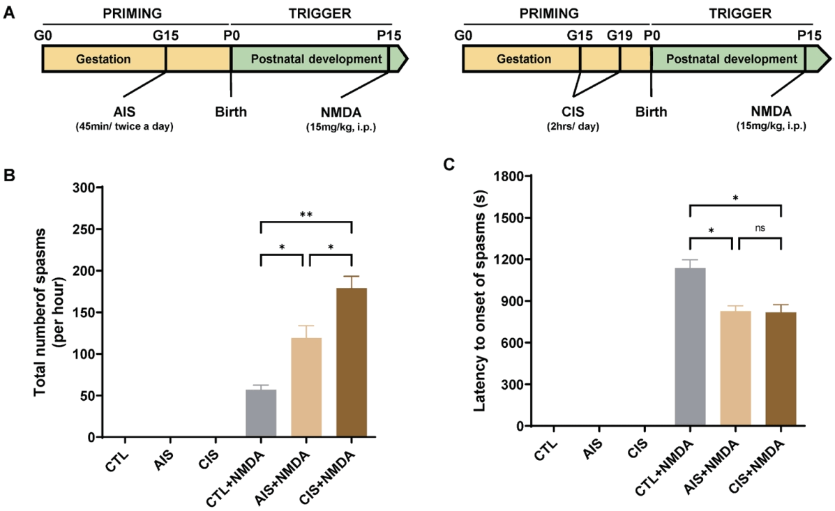
Prenatal immobilization stress priming enhances NMDA-induced spasm-like seizures. (A) Schematic diagrams illustrating the experimental timelines for the acute immobilization stress (AIS) and chronic immobilization stress (CIS) priming models. Infantile spasms were triggered in pups on postnatal day 15 by i.p. injection of NMDA (15 mg/kg). (B) Total number of spasms per hour following the NMDA challenge. Both the AIS+NMDA and CIS+NMDA groups exhibited significantly more spasms than the CTL+NMDA group, with the CIS+NMDA group showing the greatest increase. Data are presented as mean±standard error of the mean (SEM). *P=0.0289 CTL+NMDA vs. AIS+NMDA, *P=0.0331 AIS+NMDA vs. CIS+NMDA, **P=0.0011 CTL+NMDA vs. CIS+NMDA. (C) Latency to onset of spasms after NMDA injection. The CTL+NMDA group showed the longest latency, which was significantly shorter than that in the AIS+NMDA and CIS+NMDA groups. Data are presented as mean±SEM. *P=0.013 CTL+NMDA vs. AIS+NMDA, *P=0.011 CTL+NMDA vs. CIS+NMDA. AIS, acute immobilization stress; CIS, chronic immobilization stress; CTL, control; G, gestational day; i.p., intraperitoneal; NMDA, N-methyl-D-aspartate; P, postnatal day; ns, not significant.
For CIS, the protocol was as follows: pregnant rats were exposed to CIS once daily for 2 hours from gestational days 15–19 [18]. To prevent habituation to daily exposure, the restraint period was randomly scheduled at different times of day (09:00 AM–18:00 PM). For immobilization stress, a head-fixed restrainer (#JD-R-14 60×180 mm; Jeung Do Bio & Plant Co., LTD, Korea) was used (Fig. 1A).
Fifteen postnatal day 15 (P15) Sprague-Dawley rats were allocated to 3 groups: (1) control (n=5), (2) AIS offspring (n=5), and (3) CIS offspring (n=5) receiving 15 mg/kg NMDA on P15. After NMDA administration intraperitoneally, offspring in each group were monitored for 75 minutes. Spasm onset was defined as marked flexion (i.e., forelimb and trunk flexion, and flexion of the forelimbs, hind limbs, and hips). Latency from NMDA administration to spasm onset and the total number of spasms during the observation period was recorded.
2. Study measurements
1) Immunohistochemistry
Five animals per group were anesthetized with sodium pentobarbital (50 mg/kg, intraperitoneal) and perfused transcardially with heparinized phosphate-buffered saline (PBS), followed by 4% paraformaldehyde in PBS. Each brain was removed, immersed overnight in the same fixative, and cryoprotected in 10%, 20%, and 30% sucrose solutions. The brains were then embedded in Frozen Section Compound (#3801480; Leica Microsystems, Germany) and promptly frozen in 2-methylbutane, precooled to its freezing point with liquid nitrogen. Frozen coronal sections (35 μm thick) were cut using a Leica cryostat (Leica Microsystems). Parallel free-floating sections were subjected to endogenous peroxidase blocking with 1% H2O2 in PBS, followed by treatment with blocking buffer (1% fetal bovine serum in PBS and 0.3% Triton X-100) for 30 minutes and incubation with primary antibodies against glutamic acid decarboxylase isoform 67 (glutamate decarboxylase 67 [GAD67], 1:400; #MAB5406, Millipore, USA), glial fibrillary acidic protein (GFAP, 1:1000; #Z0334, Dako, USA), and ionized calcium-binding adapter molecule 1 (IBA1, 1:1000; # 019-19741, Wako Pure Chemical Industries, Japan). Immunohistochemical staining for GAD67, GFAP, and IBA1 was performed using the avidin–biotin peroxidase complex method, as described previously [19,20]. After washing with PBS, the sections were incubated with biotinylated anti-rabbit/mouse IgG and streptavidin peroxidase complexes (Vector Laboratories, USA). Immunostaining was visualized using diaminobenzidine and mounted with Polymount (Polysciences Inc., USA).
2) Sholl analysis
Sholl analysis was performed to quantitatively assess cell morphological complexity. Images of labeled cells were acquired using a bright-field microscope (Nikon) under identical acquisition settings across all experimental groups. For subsequent image processing, maximum-intensity projections were generated in ImageJ software (National Institutes of Health, USA).
3) Quantitative RT- PCR
Rat cortical tissue was harvested and homogenized for RNA extraction. Total RNA was isolated using TRIzol reagent according to the manufacturer’s instructions (GeneAll, RoboEx, Thermo Fisher Scientific, USA). RNA concentration and purity were measured using a NanoDrop spectrophotometer (Thermo Fisher Scientific). Complementary DNA (cDNA) was synthesized from total RNA using a cDNA synthesis kit (Enzynomics, Korea). Quantitative real-time polymerase chain reaction (qRT-PCR) was performed using the AriaMx Real-Time PCR system to evaluate gene expression levels. The primer sequences used were as follows: rGAPDH (5′-CTCATGACCACAGTCCATGC-3′; 5′-TTCAGCTCTGGGATGACCTT-3′), rTNF-α (5′-AGATGTGGAACTGGCAGAGG-3′; 5′-CCCATTTGGGAACTTCTCCT-3′), rIL-1β (5′-CAGCAGCATCTCGACAAGAG-3′; 5′-TGATGTGCTGCTGCGAGATT-3′), rIL-6 (5′-TGAGAAAAGAGTTGTGCAATGG-3′; 5′-CATCATCCCACGAGTCACAG-3′), riNOS (5′-AGGGAGTGTTGTTCCAGGTG-3′; 5′-GACAGATCGCCAAAACATAG-3′), rCOX-2 (5′-CAGTATCAGAACCGCATTGCC-3′; 5′-GAGCAAGTCCGTGTTCAAGGA-3′), For qRTPCR normalization, rGAPDH was used for mRNA normalization. Relative gene expression levels were calculated using the 2^−ΔΔCt method, as previously described [21].
3. Data analysis
The NIH Image program (ImageJ) was used to quantitatively assess immunohistochemical signals by densitometric measurement. Data distribution was assessed for normality before parametric testing. Quantitative data among the CTL, AIS, and CIS groups were analyzed using 1-way analysis of variance, with post hoc pairwise comparisons (including AIS vs. CIS) performed using the Tukey multiple comparisons test; comparisons between the 2 groups were performed using an unpaired Student t test. Statistical significance was set at P<0.05, and exact P values are reported in the Results and figure legends where possible. All statistical analyses were performed using the Prism 9.0 program (GraphPad Software, USA). Because statistical modeling at the offspring level—specifically, a mixed-effects model that includes the clutch as a random effect—was used, the possibility of pseudoreplication due to the clutch cannot be completely excluded.
Results
1. CIS increases susceptibility to NMDA-induced spasms in prenatally exposed rats
To investigate whether prenatal stress priming affects susceptibility to NMDA-induced spasm-like seizures, we established 2 animal models: an AIS model and a CIS model. After NMDA administration, the spasm phenotype observed in rats on postnatal day 15 (P15) was characterized by continuous tail twisting and marked flexion. In P15 rats exposed to prenatal stress, NMDA-triggered spasmlike events occurred earlier and were more frequent and intense (Fig. 1B and C). The frequency of spasms (mean± standard error of the mean [SEM]) was significantly increased in infant rats exposed to either prenatal AIS (119.0±15.0) or CIS (179.3±24.9) compared with rats in the control group (57.0±9.5) (Fig. 1B). Additionally, the latency to spasm onset (mean±SEM, seconds) was significantly decreased in rats exposed prenatally to AIS (826.7±67.3) or CIS (817.0±97.5) compared with controls (1,205.3±170.4) (Fig. 1C). These findings indicate that prenatal immobilization stress, whether acute or chronic, lowers the threshold for NMDA-induced spasm-like seizures in infant rats.
2. Prenatal immobilization stress reduces GAD67 expression in the cortex
To examine whether increased spasm susceptibility was associated with alterations in GABAergic signaling, we performed immunohistochemistry for GAD67, a key enzyme in GABA synthesis, in the cerebral cortex; the cortex was selected for this analysis because it is a principal site of GAD67-expressing interneurons and was the region used in our prior AIS model [10]. Representative photomicrographs at low and high magnification showed an apparent reduction in GAD67 immunoreactivity in the AIS and CIS groups compared with the control (CTL) group (Fig. 2A; panels A–F). Quantitative analysis of optical density values confirmed a significant decrease in GAD67 immunoreactivity in the AIS and CIS groups compared with CTL (Fig. 2G). Because optical density reflects immunoreactivity rather than interneuron number, these results suggest that prenatal immobilization stress is associated with reduced GAD67 immunoreactivity, indicating impaired GABAergic signaling or reduced GABA synthesis capacity, rather than establishing a loss of GABAergic interneurons.
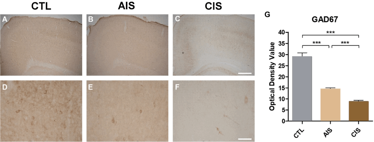
Prenatal immobilization stress reduces cortical GAD67 expression. (A–F) Representative photomicrographs of GAD67 immunohistochemistry in the cerebral cortex of rats in the CTL (A and D), AIS (B and E), and CIS (C and F) groups. Low-magnification images (A–C) showing the overall laminar distribution of GAD67-immunoreactive neurons, whereas high-magnification images (D–F) show the morphology of individual interneurons. Scale bars: 200 μm (C) and 50 μm (F). (G) Quantification of GAD67 optical density values across the groups. GAD67 immunoreactivity progressively decreased in AIS and CIS animals compared to that in CTL animals. Data are presented as mean±standard error of the mean. ***P<0.001. AIS, acute immobilization stress; CIS, chronic immobilization stress; CTL, control; GAD67, glutamic acid decarboxylase isoform 67.
3. Prenatal immobilization stress promotes neuroinflammation via astrocyte and microglial activation
Given the observed changes in GAD67 immunoreactivity, we investigated whether neuroinflammatory processes were also engaged, using markers for astrocytes (GFAP) and microglia (Iba-1). Glial activation was assessed in the hippocampus because this region is highly susceptible to NMDA-induced excitotoxic injury and glial reactivity; we acknowledge that this differs from the cortical region used for the GAD67 and qRT-PCR analyses, and interpretation of the relationship between cortical GAD67 reduction and hippocampal glial activation should therefore be considered region-specific rather than as evidence of a single unified mechanism. Immunohistochemistry of hippocampal sections revealed morphological changes in glial cells across the experimental groups (Fig. 3A–F). Among the 2 groups subjected to fetal stress, only the CIS group showed significantly higher GFAP expression in the P15 hippocampal CA3 region compared to the control group (Fig. 3G).
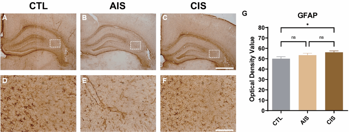
Prenatal immobilization stress induces astrocyte activation in the hippocampus. (A–F) Representative photomicrographs of GFAP immunohistochemistry in hippocampal sections from CTL (A and D), AIS (B and E), and CIS (C and F) rats. Low-magnification images (A–C) showing the overall GFAP distribution across hippocampal subregions; boxed areas are shown at higher magnifications (D–F). Scale bars, 500 μm in (C) and 100 μm in (F). (G) Quantification of the GFAP optical density in the hippocampus. The CIS group showed a significant increase in GFAP immunoreactivity compared to the CTL group, which is indicative of reactive astrogliosis. Data are presented as mean±standard error of the mean. *P=0.0476, CTL vs. CIS. AIS, acute immobilization stress; CIS, chronic immobilization stress; CTL, control; GFAP, glial fibrillary acidic protein; ns, not significant.
Analysis of Iba-1 immunoreactivity indicated a more pronounced inflammatory response. Both the AIS and CIS groups displayed significantly elevated Iba-1 optical density values compared with CTL (Fig. 4A–F). High-magnification images showed that microglia in the AIS and CIS groups exhibited an activated, bushy morphology, with enlarged cell bodies and thickened processes, consistent with a reactive microglial phenotype. Collectively, these findings suggest that prenatal immobilization stress triggers sustained neuroinflammation, characterized by both astrogliosis and microglial activation (Fig. 4G).
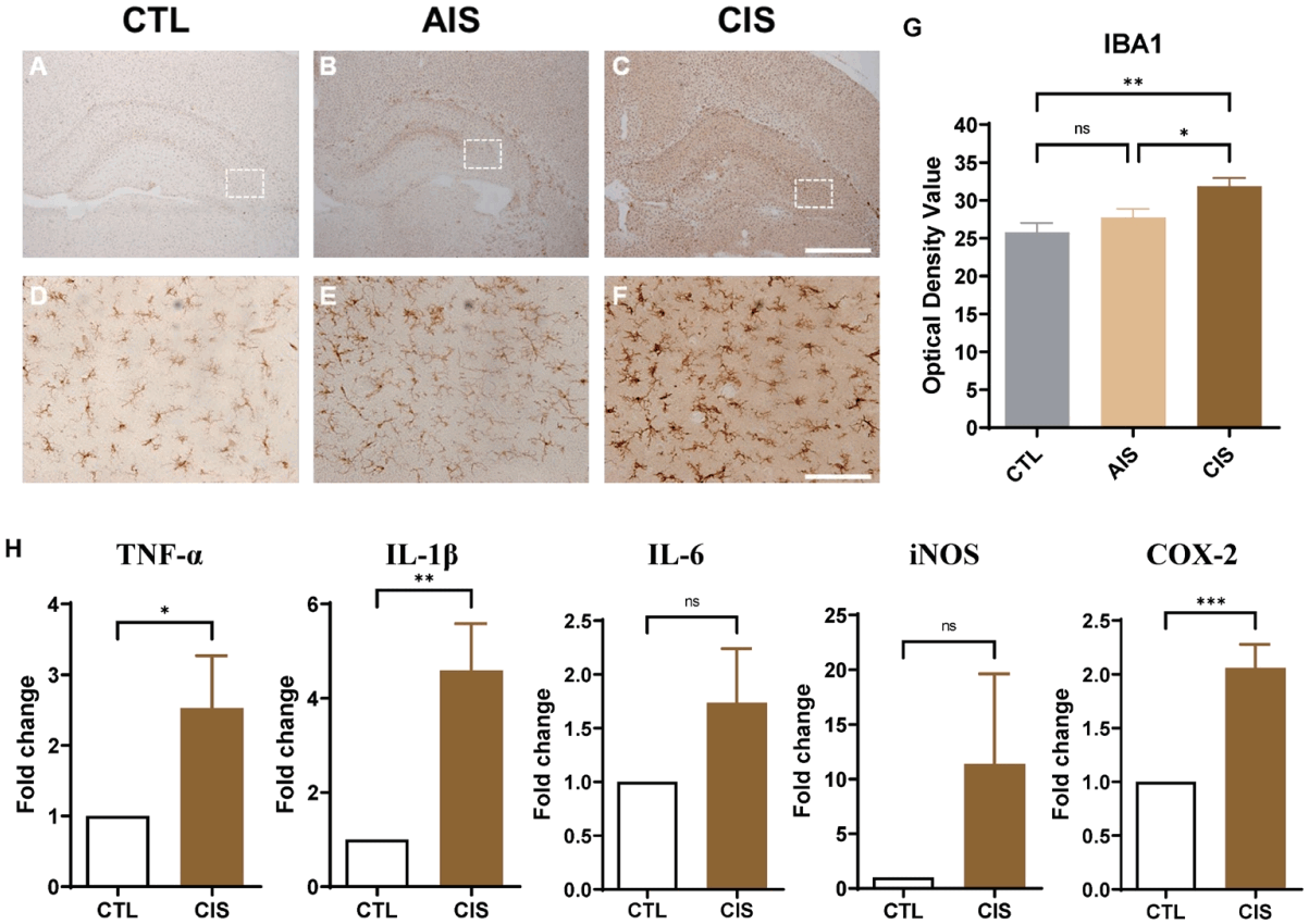
Prenatal immobilization stress activates microglia and upregulates proinflammatory mediators. (A–F) Representative photomicrographs showing IBA1 immunohistochemistry in the hippocampus of CTL (A and D), AIS (B and E), and CIS (C and F) rats. Low-magnification images (A–C) showing the regional distribution of Iba-1-positive microglia, and boxed areas are presented at higher magnifications in panels (D–F). The progressively larger soma and thickened processes observed in AIS and CIS animals were consistent with microglial activation. Scale bars: 500 μm (C) and 50 μm (F). (G) Quantification of Iba-1 optical density. The AIS and CIS groups showed significantly elevated Iba-1 immunoreactivity compared with the CTL group. Data are presented as mean±standard error of the mean (SEM). **P=0.0015, CTL vs. CIS; *P=0.0383, AIS vs. CIS. (H) mRNA expression of proinflammatory mediators—TNF-α, IL-1β, IL-6, iNOS, and COX-2—in the brains of CTL and CIS animals, assessed by quantitative real-time polymerase chain reaction. Expression levels are shown as fold changes relative to CTL. TNF-α, IL-1β, and COX-2 were significantly upregulated in CIS animals. Data are presented as mean±SEM. TNF-α, *P=0.0471; IL- 1β, **P=0.0024; and COX-2, ***P=0.0004 (unpaired Student t test). AIS, acute immobilization stress; CIS, chronic immobilization stress; COX-2, cyclooxygenase-2; CTL, control; IBA1, ionized calcium-binding adapter molecule 1; IL-1β, IL-1 beta; IL-6, IL-6; iNOS, inducible nitric oxide synthase; TNF-α, tumor necrosis factor alpha; ns, not significant.
To further characterize the neuroinflammatory profile induced by chronic prenatal stress, we quantified mRNA levels of the proinflammatory mediators TNF-α, IL-1β, IL-6, iNOS, and COX-2 in brain tissue from CTL and CIS animals by quantitative PCR (Fig. 4H).
The CIS group exhibited significantly elevated expressions of TNF-α (~2.3-fold) and IL-1β (~4.6-fold) compared with CTL animals. IL-6 and iNOS expression showed trends toward upregulation in the CIS group, although these differences did not reach statistical significance, likely because of high interindividual variability. COX-2 mRNA levels also increased significantly in the CIS group (~2.1-fold), indicating enhanced inflammatory signaling.
4. Prenatal immobilization stress differentially affects microglial morphological complexity
To characterize microglial morphology following prenatal inflammatory priming and NMDA-triggered seizure induction, we conducted Sholl analysis of Iba-1-immunolabeled microglia in the cerebral cortex. Concentric circles were drawn at 1-μm intervals from the center of each cell body, and process intersections were counted at each radius to generate an intersection-distance profile for each group (Fig. 5A).

Prenatal immobilization stress differentially alters microglial morphological complexity after NMDA challenge. (A) Representative Sholl analysis images of IBA1-immunolabeled microglia from CTL, AIS, and CIS animals. Concentric circles were drawn at 1-μm intervals from the center of the cell body, and white traces indicate individual microglial processes. The AIS microglia display more extensively ramified arbors that extend to greater radial distances than those of the CTL and CIS cells. (B) Sholl intersection profiles of IBA1-positive microglia in the CTL-NMDA, AIS-NMDA, and CIS-NMDA groups. The number of process intersections is plotted as a function of distance from the cell body (μm). The AIS-NMDA group exhibited a markedly broader, higher-intersection curve with a peak of ~6–7 intersections at 7–8 μm and detectable arborization extending to ~22 μm. In contrast, the CTL-NMDA and CIS-NMDA groups display similar, more compact profiles, peaking at ~5 intersections near 5 μm and declining sharply beyond 12 μm. Data are presented as mean±standard error of the mean. ***P<0.0001, **P=0.0025 (11.5060 μm), 0.0036 (14.2455 μm), 0.0029 (16.9860 μm) (CTLNMDA vs. AIS-NMDA). AIS, acute immobilization stress; CIS, chronic immobilization stress; CTL, control; IBA1, ionized calcium-binding adapter molecule 1; NMDA, N-methyl-D-aspartate.
Representative Sholl overlay images (Fig. 5A) show clear group differences in the spatial distribution of microglial processes in the CTL, AIS, and CIS groups. In CTL animals, microglia display relatively sparse, short processes that intersect only the innermost concentric rings. Conversely, AIS microglia exhibited substantially more ramified arbors, with processes extending well beyond the intermediate radii. CIS microglia showed an intermediate morphology compared with AIS microglia, yet appeared more ramified than CTL cells, consistent with a graded inflammatory priming effect.
Quantitative Sholl analysis confirmed these observations (Fig. 5B). In the CTL-NMDA group, the intersection curve peaked at approximately 5 intersections at about 4 μm from the cell body and then declined steeply, reaching near-zero values beyond 12 μm. The significantly greater area under the intersection-distance curve in AIS-NMDA animals indicates that acute prenatal immobilization stress promotes a more highly ramified, surveillance microglial phenotype, even after an acute NMDA challenge.
In contrast, the CIS-NMDA group exhibited an intersection profile that closely mirrored that of the CTL-NMDA group, peaking at approximately 4–5 intersections near 4–5 μm and showing a similarly rapid decline beyond 10 μm. This convergence between the CIS-NMDA and CTL-NMDA profiles indicates that, unlike AIS, chronic prenatal stress does not preserve the more highly ramified, surveillance-like microglial morphology after NMDA challenge. However, because the CTL group also received NMDA, this shared deramified profile most likely reflects, at least in part, NMDA-induced acute excitotoxicity common to both groups, rather than a CIS-specific effect.
Discussion
In this study, we investigated whether prenatal immobilization stress, particularly CIS, increases susceptibility to NMDA-induced spasm-like seizures in infant rats and examined associated changes in GABAergic signaling and glial activation. Our main findings can be summarized as follows: (1) chronic prenatal stress lowered the threshold for NMDA-induced spasm-like seizures, producing a greater increase in spasm frequency than acute prenatal stress (AIS); (2) prenatal stress, whether acute or chronic, was associated with reduced cortical GAD67 immunoreactivity; (3) prenatal stress was associated with astrocyte and microglial activation and upregulation of proinflammatory mediators after NMDA challenge; (4) acute and chronic prenatal stress appeared to have distinct, rather than simply graded, biological consequences, as reflected by differing microglial morphological responses between the AIS and CIS groups; and (5) several methodological limitations, discussed below, temper the interpretation of these findings. It should be noted that although the CIS group showed the greatest increase in spasm frequency, the AIS and CIS groups did not differ consistently across all outcome measures, suggesting that acute and chronic prenatal stress may engage both overlapping and distinct mechanisms rather than a simple linear dose-response relationship.
IESS are a form of developmental and epileptic encephalopathy that occurs in early childhood. In many cases, patients do not receive adequate medical therapy, which is usually responsible for the observed developmental delay. Understanding the cryptogenic etiology of this condition remains challenging [2-4]. However, various hypotheses are currently being explored. Determining the cause of a seizure is important for defining the treatment target. Thus, studies examining the mechanisms underlying IESS also contribute to research aimed at understanding developmental delays.
In our study, prenatal exposure to CIS, coupled with postnatal NMDA-triggered spasm-like seizure induction, increased spasm frequency and decreased spasm latency compared with control conditions, with a greater increase observed in the CIS group than in the AIS group for spasm frequency. Prior work using the AIS model reported decreased KCC2 expression and increased intracellular Cl− concentration in GABAergic neurons, together with a depolarized resting membrane potential, and proposed that these changes could lower the threshold for spasm-like seizures [10]. KCC2 expression and membrane potential were not directly assessed in the present study; therefore, the reduced cortical GAD67 immunoreactivity we observed in the CIS group should be regarded as indirect, associative support for—rather than direct confirmation of—the hypothesis that disrupted chloride homeostasis and impaired GABAergic inhibition contribute to prenatal stress-related spasm susceptibility. Direct assessment of KCC2 expression and chloride handling in the CIS model, together with dose-response characterization of the stress exposure required to alter this pathway, will be necessary in future studies to test this hypothesis.
Consistent with the greater spasm frequency observed in the CIS group, several clinical studies similarly report that chronic stress has a more pronounced adverse effect on seizures than acute stress. Psychological stress is consistently identified as the most frequently self-reported seizure precipitant among patients with epilepsy. However, clinical evidence demonstrates that its effects on seizure severity and frequency are not uniform and that chronic stress exerts more pronounced and sustained adverse effects than acute stress [11]. A prospective diary study in patients with localization-related epilepsy demonstrated that incrementally higher levels of sustained self-reported stress and anxiety were significantly associated with an increased probability of seizure occurrence on the following day (odds ratios: 1.06 and 1.07 per unit increase), supporting the dose-dependent, cumulative nature of chronic stress on seizure susceptibility [22]. In a controlled clinical study of 64 children with epilepsy using the Trier Social Stress Test and a 6-week seizure-stress diary, children whose seizures were triggered by acute stress showed a paradoxically blunted cortisol response to acute psychosocial stimuli compared to the control group [12]. Direct human tissue evidence from surgical specimens of patients with temporal lobe epilepsy has demonstrated that chronic glucocorticoid receptor activation, consistent with chronic stress, impairs long-term potentiation in the human temporal cortex. This mechanism is distinct from the transient modulatory effects of acute glucocorticoid exposure, suggesting that chronic stress independently undermines synaptic homeostasis, thereby exacerbating epilepsy [23].
A key limitation of this study is that spasm-like events were identified based on behavioral observation alone, without electroencephalographic (EEG) confirmation, and the animals did not exhibit spontaneous seizures; the NMDA-induced events described here should therefore be regarded as a pharmacologically provoked spasmlike model rather than a full electroclinical correlate of IESS. Other studies using similar models have reported EEG findings and drug-response profiles consistent with IESS [10,24].
This study has several limitations that should be acknowledged. First, each group (CTL, AIS, and CIS) consisted of only 5 animals, and sample size was not derived from a formal a priori power calculation; the present findings should therefore be regarded as preliminary, and adequately powered replication studies with larger cohorts are warranted. Second, although we report the number of offspring analyzed per group, we did not clearly report the number of dams per group or explicitly model litter as a random effect; because pups from the same litter are not statistically independent, this raises the possibility of pseudoreplication, and future studies should report dam and litter numbers and consider litter-based statistical models. Third, this study did not include no- NMDA control groups—that is, CTL, AIS, and CIS animals that were not challenged with NMDA and were not monitored over an extended baseline period. As a result, we cannot fully exclude the possibility that spontaneous epileptiform activity could occur independently of NMDA, nor can we fully distinguish the direct, baseline effects of prenatal stress from NMDA-induced excitotoxic or postictal changes in GAD67 immunoreactivity and glial activation; consequently, our mechanistic interpretations should be tempered accordingly, and future studies incorporating nonchallenged, stress-only cohorts with continuous long-term monitoring would help clarify the independent contribution of prenatal stress alone. Fourth, qRT-PCR analysis of proinflammatory mediators (Fig. 4H) was performed only in CTL and CIS animals; the AIS group was not included, so it remains unknown whether AIS produces similar transcriptional changes. Fifth, although the CIS group showed the greatest increase in spasm frequency, the AIS and CIS groups did not differ consistently across all outcome measures—for example, in microglial morphological complexity after NMDA challenge, AIS microglia retained a more ramified, surveillance-like morphology, whereas CIS microglia showed a more deramified, activated profile similar to that seen after NMDA challenge alone. This distinction may reflect different microglial priming states following acute versus chronic prenatal stress (a more transient, adaptive response after a single stressor versus a more persistently reactive phenotype after repeated stress), but given the limitations noted above, this interpretation remains speculative and suggests that acute and chronic prenatal stress may not simply produce graded effects of differing magnitude. Sixth, neuroinflammatory changes were examined only at P15, the developmentally validated age for this NMDA-induced infantile spasms model; we therefore cannot determine whether these effects are transient or persist beyond this age-specific window, and future studies comparing multiple postnatal ages would help clarify their developmental specificity. Finally, the associations observed among neuroinflammation, reduced GAD67 immunoreactivity, and increased spasm susceptibility do not establish causality; interventional studies using anti-inflammatory treatments or GABAergic rescue approaches, together with no-NMDA controls, will be required to test whether these changes causally contribute to the phenotype.
Although sex differences in seizure susceptibility are well established in adult rodents, this dimorphism is largely absent at P15. Immature rats show no sex-specific differences in acute seizure susceptibility, unlike adults [22], consistent with estradiol manipulation having no effect on spasms in this model around the same age [23]. This likely reflects the fact that sex differences in seizure susceptibility depend on gonadal hormones that intensify only after puberty [24], a window P15 precedes. We note that regional KCC2 dimorphism has been reported at P15 [25], but this does not appear to produce a measurable wholeanimal difference in spasm susceptibility, so we did not stratify our analyses by sex.
Although AIS has previously been associated with seizures, the relevance of CIS to seizures remains uncertain. Ahmadzadeh et al. [26] reported that short-term prenatal stress increased susceptibility to pilocarpine-induced seizures. Many studies have reported that chronic prenatal stress is associated with depression [27], memory impairment [28], and even schizophrenia [8]. However, the relationship between CIS and seizures remains unclear. Here, we found that chronic prenatal stress increased seizure susceptibility in the offspring.
In conclusion, our findings indicate that prenatal stress, particularly CIS, increases susceptibility to NMDA-induced spasm-like seizures in infant rats and is associated with reduced GAD67 immunoreactivity and enhanced glial activation. Because the present data demonstrate associations rather than causal relationships, future studies incorporating no-NMDA tissue controls, adequately powered litter-based designs, EEG confirmation, and interventional approaches such as anti-inflammatory or GABAergic rescue strategies will be needed to determine whether neuroinflammation and GABAergic dysfunction causally contribute to this phenotype.
Notes
Conflicts of interest
No potential conflict of interest relevant to this article was reported.
Funding
All authors gratefully acknowledge financial support from the National Research Foundation of Korea (NRF), funded by the Ministry of Science and ICT, Republic of Korea (2022R1A2C1007320, RS-2025–16067883). This work was also supported by a research fund from Chungnam National University (Grant No. 2023–0586–01). This research was supported by the ANCHOR program through the Daejeon ANCHOR Center, funded by the Ministry of Education (MOE) and Daejeon Metropolitan City, Republic of Korea (2026- ANCHOR-06–011).
Acknowledgments
We thank the use of the Slide scanner Core Facility of the Regional Medical Research Capability Enhancement Project, Biomedical Research Institute, Chungnam National University Hospital, for providing technical assistance, instrumentation, and data analysis support.
Author contribution
Conceptualization: JWK; Formal Analysis: HHK, SGC; Data Curation: SB; Writing–Original Draft: HJS, JWK; Writing – Review & Editing: HJS, JWK