Usefulness of anterior uveitis as an additional tool for diagnosing incomplete Kawasaki disease
Article information
Abstract
Purpose
There are no specific tests for diagnosing Kawasaki disease (KD). Additional diagnostic criteria are needed to prevent the delayed diagnosis of incomplete Kawasaki disease (IKD). This study compared the frequency of coronary artery lesions (CALs) in IKD patients with and without anterior uveitis (AU) and elucidated whether the finding of AU supported the diagnosis of IKD.
Methods
This study enrolled patients diagnosed with IKD at The Catholic University of Korea, Uijeongbu St. Mary's Hospital from January 2010 to December 2014. The patients were divided into 2 groups: group 1 included patients with IKD having AU; and group 2 included patients with IKD without AU. We analyzed the demographic and clinical data (age, gender, duration of fever, and the number of diagnostic criteria), laboratory results, and echocardiographic findings.
Results
Of 111 patients with IKD, 41 had uveitis (36.98%, group 1) and 70 did not (63.02%, group 2). Patients in group 1 had received a diagnosis and treatment earlier, and had fewer CALs (3 of 41, 1.7%) than those in group 2 (20 of 70, 28.5%) (P=0.008). All 3 patients with CALs in group 1 had coronary dilatation, while patients with CALs in group 2 had CALs ranging from coronary dilatation to giant aneurysm.
Conclusion
The diagnosis of IKD is challenging but can be supported by the presence of features such as AU. Group 1 had a lower risk of coronary artery disease than group 2. Therefore, the presence of AU is helpful in the early diagnosis and treatment of IKD and can be used as an additional diagnostic tool.
Introduction
Kawasaki disease (KD) is an acute, systemic vasculitis of unknown etiology that occurs predominantly in infants and young children less than 5 years old. It was firstly reported by Kawasaki in 19671), and has subsequently been reported worldwide in children of all races. A delayed diagnosis of KD, especially incomplete Kawasaki disease (IKD)2); which has persistent fever with fewer than four other principal clinical features of KD, can lead to the development of coronary artery lesions (CALs). Recently, the diagnosis of IKD has been increasing, and the reported prevalence was 15% to 36.2% among patients with KD345678), making it difficult for pediatrician to diagnose KD. Therefore, additional clues that are helpful in the diagnosis of IKD are needed for the early detection and treatment of IKD. Suggested clues are erythema and induration of a previous Bacille Calmette-Guerin inoculation site910), anterior uveitis (AU)11), elevated serum levels of alanine aminotransferase (ALT)12), brain natriuretic peptide (BNP)13) and N-terminal pro-BNP1415), hyponatremia16), echocardiographic abnormalities other than CALs17) (pericardial effusion, dysfunction of the left ventricle, mitral regurgitation). AU is a common finding in the early stage of KD, so a slit lamp examination may be a helpful in the early detection of patients with IKD. No study has examined the usefulness of AU for the early recognition and treatment of IKD in Korea. Therefore, we retrospectively investigated the diagnostic value of AU for the early detection of IKD.
Materials and methods
1. Patients
This study enrolled patients diagnosed with KD at The Catholic University of Korea, Uijeongbu St. Mary's Hospital from January 2010 to December 2014. The patients were divided to 2 groups on admission: group 1 presented with sustained fever and two or three principal features of KD with AU; group 2 presented with sustained fever with two or three principal features of KD without AU. We analyzed the following data obtained from a retrospective review of the charts of the enrolled patients with KD during hospital admission leading to the final diagnosis: clinical and demographic data (age, gender, duration of fever, and the number of diagnostic criteria), laboratory values before the administration of intravenous immunoglobulin (IVIG), response to IVIG, and echocardiographic findings. All patients were treated with IVIG (2 g/kg) and aspirin (50 mg/kg) as the first-line treatment. The response to the initial IVIG treatment was considered "refractory"12) if persistent or recrudescent fever was noted at least 36 hours after IVIG administration. Refractory KD was treated using additional IVIG infusions or the second-line therapy, such as systemic corticosteroids or infliximab.
2. Ophthalmologic examination
All patients with IKD underwent ophthalmology consultations to confirm the existence of AU. AU was diagnosed by slit-lamp examination and graded using Standardization of Uveitis Nomenclature (SUN) Grading Scheme for Anterior Chamber Cells and SUN Grading Scheme for Anterior Chamber Flare.
3. Blood tests
The laboratory tests were performed on admission (before the IVIG infusion) and during the course of the disease, including compete blood cell counts, erythrocyte sedimentation rate, C-reactive protein, serum albumin, and serum ALT.
4. Echocardiographic examinations
Two-dimensional echocardiography was performed during hospitalization and follow-up periods at the outpatient clinic (1 month and 1 year after discharge). The internal diameters of left main, left anterior descending, and right coronary arteries were measured. The normal ranges for coronary artery (CA) size defined according to the age or body weight were used18). In children younger than 5 years, an internal luminal diameter (ILD) ≤3.0 mm was considered normal, while in children aged 5 years or older, an ILD≤4.0 mm is considered normal. Grouping children by weighing less than 12.5, 12.5–27.5, and more than 27.5 kg, the normal ranges of ILD were defined as ≤2.5, and 2.5–3.0, and 3.0–5.0 mm, respectively. If the ILD of CA segment was enlarged less than 1.5 times of the upper normal limit, it was defined as dilatation, and if the ILD was enlarged 1.5 times or more, it was defined as aneurysm.
5. Statistical analysis
All statistical analyses were performed using IBM SPSS ver. 18.0 (IBM Co., Armonk, NY, USA), and the data were presented as mean±standard deviation or as percentages. Variables were compared between 2 groups using the unpaired Student t test, Pearson chi-square test, and one-way analysis of variance. P values of <0.05 were considered statistically significant. This study was approved by the Institutional Review Board of the School of Medicine of The Catholic University of Korea (UC15RISI0098).
Results
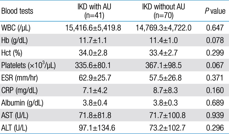
During the study period, 111 patients were diagnosed having IKD at Uijeongbu St. Mary's Hospital. Of these, 41 had AU on admission (group 1), and 70 did not have AU (group 2). The demographic data of each group are summarized in Table 1. The mean age was significantly older in group 1 than in groups 2 (46.5±19.93 months vs. 29.8±21.28 months, P<0.001). This data might mean that there could be some patients with AU in the group 2, because the very young aged patients might not get the correct diagnosis of AU due to the lack of cooperation during slit lamp examination. Males predominated (67 of 111, 60.3%) as like as the patients with complete KD and the male-to-female ratio was 1.5:1. The duration of fever before the first IVIG was not significantly different between the 2 groups (P=0.533). There was no significant difference in the number of diagnostic criteria between 2 groups (Table 2), and all of the enrolled patients had desquamation of finger tips and thrombocytosis in the subacute phase of KD, confirming the final diagnosis of KD. The rates of additional IVIG treatment did not significantly differ between the two groups. The necessity of second-line therapy, such as systemic corticosteroid or infliximab, was significantly higher in group 2 than in group 1. Group 2 had a significantly longer duration of hospitalization than group 1. The laboratory results on admission did not differ significantly between the two groups (Table 3). CA dilatation was observed in 3 patients in group 1 (1.7%), 19 patients in group 2 (27.1%), and the rate of dilatation was significantly higher in group 2 than in group 1 (Table 4) (P=0.008). The only 1 patient in group 2 (1.4%), who received infliximab, developed giant coronary aneurysm. The fact that group 1 had lower risk of CALs than group 2 seemed to demonstrate that the detection of AU could lead to earlier diagnosis and treatment of patients with IKD before they develop CALs (relative risk [RR], 1.298; 95% confidential interval, 10.93–1.540).

Laboratory results of patients with incomplete Kawasaki disease with and without anterior uveitis at admission
Discussion
Kawasaki disease is the most common acquired heart disease in children. Delayed treatment can cause cardiac complications, including CA dilatation and aneurysm, myocardial infarction, pericardial effusion, valvular regurgitation. KD is diagnosed using the diagnostic criteria1218) based on the clinical symptoms and laboratory findings, because there is no specific diagnostic test. Ophthalmologic manifestations of KD have been described since 198119), mostly nonexudative bulbar conjunctivitis and uveitis12). Superficial punctate keratitis, vitreous opacity, choroidal and retinal changes, papilledema, subconjunctival hemorrhage, and macular edema are less common20). Some studies have included AU as an additional diagnostic tool that can be helpful for the early recognition and treatment of IKD212223) and the prevention of CALs. The patients with IKD younger than 6 months of age, or older than 5 years of age, have higher possibility of developing CA complications due to late diagnosis and treatment35). Uveitis can be classified anatomically, with AU accounting for 30%–40%, posterior uveitis accounting for 40%–50%, intermediate uveitis accounting for 10%–20%, and diffuse uveitis accounting for 5%–10%24). The most common cause24) of AU in children is juvenile idiopathic arthritis, while other diseases can be associated with AU including KD, retinoblastoma, leukemia, juvenile xanthogranuloma, Coat's disease, retinitis pigmentosa, herpes virus infection, sarcoidosis, and retained intraocular foreign bodies following trauma. CALs were significantly less common in group 1 than group 2 (7.3% vs. 28.5%, P=0.008). Therefore, if a patient has an unexplained persistent fever and IKD is suspected, an ophthalmic examination is needed to identify the presence of AU. Korea has the second highest incidence rate of KD in the world. However, we do not have our own diagnostic criteria for KD and should establish Korean diagnostic guidelines of the disease with additional criteria such as AU for the early diagnosis of IKD and prevention of CA complications. To do this, it is necessary to objectively assess whether the detection of AU can help to diagnose and reduce the occurrence of CALs by preventing treatment delay through the triennial nationwide epidemiological survey in Korea. This study is the first such study in Korea, but it has some limitations. First, all of the patients with KD were not included, and only those with suspected IKD underwent ophthalmologic examinations. Second, the study had a small sample size because it was conducted at a single institute. Finally, the study was not a nationwide prospective study. We are planning a more objective, systematic study on IKD. We recommend that all patients with KD should be consulted to an ophthalmologist to determine the presence of AU. We think that AU may be recognized as one of the essential diagnostic criteria of KD, and the presence of AU may lead to the early diagnosis and treatment of IKD, reducing the occurrence of CALs. In conclusion, we recommend that pediatricians should consider AU as an important finding and cooperate with ophthalmologists when managing patients with suspected IKD.
Acknowledgment
The authors are indebted to all the contributors that participated in this survey.
Notes
Conflicts of interest: No potential conflict of interest relevant to this article was reported.


